

Custom Search
|


|
Dermpath-India Pathology of Arteriovenous Hemangioma [Arteriovenous Malformation] (2022) |
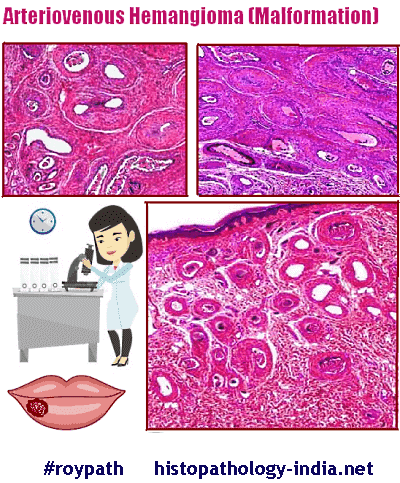
| Arteriovenous hemangioma (malformation) is a benign acquired cutaneous vascular lesion and is characterized by a complex network of intercommunicating arterial and venous structures. |
|
Arteriovenous hemangioma (malformation) is a benign acquired cutaneous
vascular lesion and is characterized by a complex network of intercommunicating arterial and
venous structures. The etiology of arteriovenous hemangioma is uncertain; endocrine and inflammatory stimuli may activate an underlying vascular malformation. Some lesions, especially those in younger patients, may be true hamartomas. Arteriovenous malformations grow proportionately with the patient; do not involute; and may sometimes increase in size because of vascular ectasia induced by conditions such as sepsis, trauma, puberty, and pregnancy. Arteriovenous hemangioma are divided into types according to the depth of involvement: 1. Deep form: May be associated with arteriovenous shunting and soft tissue hypertrophy. Age: Young adults and adolescents. Site: Commonly affect the head, neck and lower extremity. Clinical presentation: Can present with severe symptoms of heart failure or Kasabach Merritt syndrome. This form of hemangioma may be difficult to diagnose. Clinicopathologic correlation with arteriographic studies is essential in establishing the diagnosis. 2. Superficial form: (cirsoid aneurysm or acral arteriovenous tumour) Age: Middle aged and elderly adults. Site: Usually affect the head and neck region. These are often localized to the dermis and submucosa of the lips and perioral skin. Clinical presentation: Small reddish blue papule. Symptoms are mild and include pain and intermittent bleeding. Histopathological features: In both forms there are thick and thin-walled sized arteries and veins in close association with one another. Focally, some tumours resemble capillary and cavernous hemangioma. There may be focal thrombosis, secondary dystrophic calcification and mild inflammation. Serial sections are helpful in demonstrating continuities or shunts between arteries and veins. Pathologists rely on elastic stains for making a definitive diagnosis of arteriovenous malformations, because arteries and arterioles (with elastic lamina in their walls) are an integral part of Arteriovenous malformations. These lesions grow slowly and achieve only a small size and are usually cured by simple excision.
|
|
|
