

Custom Search
|

|
Infectious Disease Online Pathology of Anthrax Infection
|
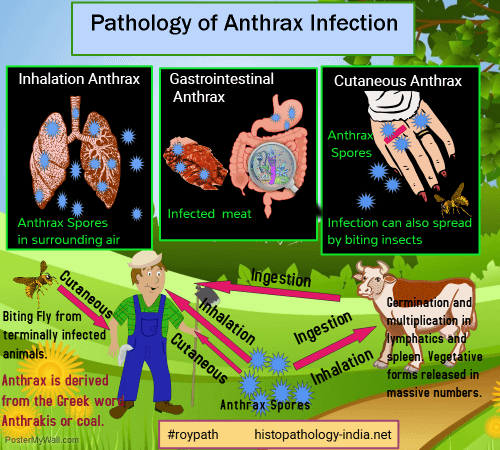
The word Anthrax is derived from the Greek word anthrakis or 'coal' in reference to the black skin lesions victims develop. Anthrax is of great historical interest for it was the first human disease of proved bacterial origin. It was described in Homer's Iliad. In 1868, Anthrax was observed under a microscope. In 1876, German bacteriologist Robert Koch confirmed bacterial origin of anthrax. Anthrax is a rare acute infectious febrile septicemic disease caused by Bacillus anthracis, a large Gram-positive, spore-bearing bacillus which normally causes disease in animals. Highly resistant spores are formed in soil and dead animals (not in diseased animal or man). Man is rarely, infected by spores, through abrasion of skin, by inhalation or by ingestion of infected meat. Symptoms start within 1-2 days of infection. Once in man, spore germinates to vegetative form and produce lethal toxins. Anthrax is an occupational disease of farmers, butchers and dealers of hides, animal hair, wool etc. Three forms of the disease: 1. Cutaneous: Malignant pustule - Hide porter's disease. This is the most common form (95%). Bacteria enters through abrasions of skin. It is commonly seen in head and neck. Initial papule is surrounded by edema and hyperemia. Papule becomes vesicle and ruptures with central necrosis forming ragged, black ulcer, surrounded by minute vesicles and papules. This painless ulcer is called an eschar. Lesion is associated with intense itching & heals within 1-2 weeks. Regional lymph nodes may be enlarged indicating progressive lesion leading to septicemia and death. Microscopic features: Epithelium is edematous with loss of continuity. Sub-epidermal, chronic and acute infiltrates involving adipose tissue and capillaries, with areas of necrosis. Gram stain shows large solid and beaded gram-positive rods, particularly beneath the epithelium. The bacilli are not really visible on the hematoxylin and eosin stain. 2. Inhalation: Pulmonary (Pneumonic) type: Wool Sorter's Disease. Inhalation of dust or filaments of wool from infected animals, particularly in wool factories. Pulmonary anthrax causes extensive serofibrinous exudate in large areas of the lung and sometimes consolidation of an entire lobe. It causes hemorrhagic bronchopneumonia, necrosis of alveolar septa and thrombi in alveolar capillaries leading to septicemia and death. Hemorrhagic meningitis may occur as a complication. 3. Gastrointestinal type: This is due to ingestion of contaminated meat. Ulceration of the stomach or bowel and invasion of regional lymphatics are usual features. The hemorrhagic gastroenteritis of anthrax may cause obstruction or perforation but death is commonly caused by electrolyte imbalance and hemoconcentration produced by fulminant diarrhea and massive ascites. Septicemia usually follows pulmonary anthrax than malignant pustule. In pulmonary antrax bacilli are phagocytosed by alveolar macrophages and transported to regional lymph nodes and then to the blood by way of the thoracic duct. Disseminated intravascular coagulation complicates septicemia. Bacterial toxin causes fatal depression of the respiratory center. Death is the usual consequence following profound systemic manifestations. Morphologic changes in Anthrax infection: It is characterized principally by a bloody mucinous edema that affects many tissues and is found in most serous cavities. Meningitis may be a prominent feature. Laboratory diagnosis: 1. Smears from pustules, sputum, CSF. ; 2. Blood culture
|
|
|

![]()
Copyright © 2022 histopathology-india.net