

Custom Search
|
|
Custom Search
|
|
Infectious Disease Online Pathology of Blastomycosis
|
Syn: North American blastomycosis.
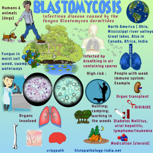
Blastomycosis is caused by Blastomyces dermatitidis. The organism is spherical, single budding, broad base with multiple basophilic nuclei in double walled central body. Epidemiology: North America and parts of Africa. Cases have been reported in India. Reservoir: These are normally present in soil . Mode of infection: Enters body by inhalation. Once inhaled in the lungs the organism multiplies and is disseminated by hematogenous route or through lymphatics to other organs. Presentation: 3 clinical forms: (i) Pulmonary Blastomycosis (ii) Primary Cutaneous form (iii) Disseminated Blastomycosis. Organs commonly involved are Lungs and Skin. Others sites include bone, joints, trachea, larynx, genitourinary organs, prostate and meninges. Lungs:
Initial pulmonary lesion is solitary or bilateral consolidation with hilar lymphadenopathy. Pulmonary lesion usually resolves by scarring, some may progress to miliary or focal consolidation with cavitation.
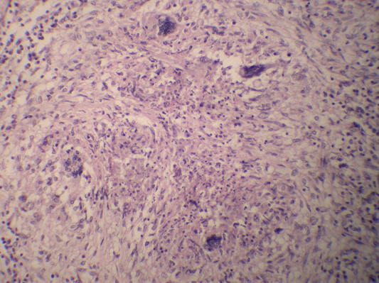
Microscopic features: Suppurative or granulomatous lesions with numerous organisms in epithelioid and giant cells or lie free in the microabscesses. Skin: Primary cutaneous Blastomycosis is rare and occurs following direct inoculation of the organism into the skin. Secondary cutaneous lesions occur in the course of disseminated disease. Skin lesions are commonly located on the face, neck and extremities. Usually presents as verrucous nodules or ulcerated plaques. Widespread pustular eruptions have been reported. Multiple lesions may appear. In some cases there is healing and scar formation.
|
|
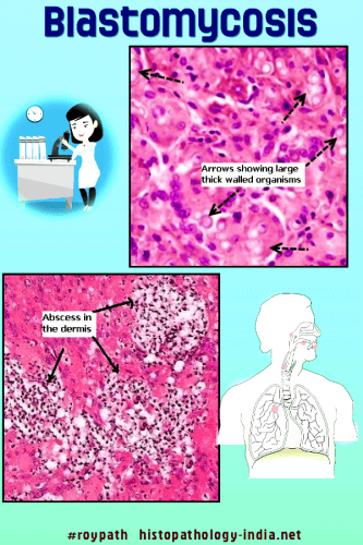
Microscopic features: A- Cutaneous lesions of disseminated blastomycosis: i) Pseudoepitheliomatous hyperplasia: Differential diagnosis- Chromomycosis & sporotrichosis. Epidermis lacks the cytological atypia seen in squamous cell carcinoma. ii) Microabscesses - Intraepidermal and in the dermis. iii) Suppurative granulomas. iv) Thick-walled yeasts present in the center of the abscesses and in giant cells. Yeasts are also present extracellularly in the dermis. V) The organisms are best demonstrated by PAS and Silver Methenamine stains. B- Primary cutaneous lesion: Epidermal hyperplasia is less prominent ; Giant cells and granulomas are usually not present ; Numerous organisms are present ; There is a mixed inflammatory infiltrate containing lymphocytes and neutrophils.
|
|
|
Visit:- Dermatopathology Online

Consultant Histopathologist (Kolkata - India)
|
![]()
Copyright © 2022 histopathology-india.net
