Custom Search
|


|
Dermpath-India Pathology of Bullous Pemphigoid Dr Sampurna Roy MD 2023
|

|
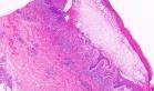
Bullous Pemphigoid was first described in 1953, is a chronic subepidermal blistering disease. Age: Most commonly occurs in elderly persons (in the seventh or eighth decade of life) but occasionally it is seen in young adults and even in children. Site: Lower part of abdomen, inner part of thighs, flexor surface of the arm and leg , axillae, groin, and palmar and plantar surfaces; oral lesions have also been reported ; Other mucosal surfaces are usually not involved. Clinical presentation: Multiple tense bulla on normal or erythematous skin of variable size. May present as erythematous macules, verrucous and vegetating lesions, urticarial plaques and crusted erosions, or annular lesions. Cause of the disease: Caused by autoantibody-mediated disruption of adhesion between basal keratocytes and the basement membrane, i.e., antibodies there bind the antigens of bullous pemphigoid. Antibodies against two hemidesmosomal components, BP antigen 2 (180 KDa) and BP antigen 1 (230 KDa), have been detected in sera of the patients. Circulating antibodies against the BP180 component are sufficient to produce a subepidermal blister. Triggering factors: Trauma ; burns ; ultraviolet irradiation, shortly after vaccination; drugs; penicillin derivatives ; antibiotics ; ibuprofen and other non-steroidal anti-inflammatory and antipsychotic drugs. May occur in association with other diseases: Example: Rheumatoid arthritis, lichen planus, psoriasis, systemic lupus erythematosus, diabetes mellitus, primary biliary cirrhosis , ulcerative colitis. etc. Microscopic features:
Early (urticarial)- Perivascular and interstitial infiltrate of lymphocytes and eosinophils ; Eosinophils scattered throughout the upper part of the reticular dermis, in the edematous papillary dermis, and along the dermoepidermal junction; Eosinophils may be present in tiny collections together with spongiosis Fully developed (vesicular) - Perivascular and interstitial infiltrate of lymphocytes and many eosinophils ; Numerous eosinophils throughout the edematous upper dermis; Subepidermal blister within which numerous eosinophils usually are present ; Dermal papillae are often preserved ; Spongiosis is often present at the sides of the blister. Late (bullous) - All the changes seen in developed fully lesions ; Sometimes there is formation of spongiotic vesicles together with edema of the papillary dermis. This feature is so prominent at times that it forms subepidermal vesiculation ; Re-epithelialization from infundibular and eccrine ductal epithelium is noted in some lesions Rarely other histological changes may be present - Cell-poor type ; An intraepidermal blister occurs in conjunction with a subepidermal blister ; Presence of an infiltrate of lymphocytes and plasma cells ; Presence of predominantly neutrophils.
Differential diagnosis of Bullous Pemphigoid: 1. Urticarial lesions of pemphigus vulgaris 2. Blisters of herpes gestationis are indistinguishable from those of cell-rich bullous pemphigoid. 3. Dermatitis Herpetiformis : Collections of neutrophils at the tip of dermal papillae and in subepidermal spaces. Nuclear "dust" of neutrophils and bands of neutrophils are noted 4. Cicatricial pemphigoid . 5. Incontinentia pigmenti in the vesicular stage. 6. Urticaria - No blisters are present consists of lymphocytes, neutrophils, and eosinophils around venules in the reticular dermis. 7. Insect Bite - a wedge-shaped infiltrate of lymphocytes and eosinophils around venules of the reticular dermis and eosinophils scattered interstitially. 8. Pruritic urticarial papules and plaques of pregnancy.
|
|
|