

Custom Search
|
|
Custom Search
|
|
Dermpath-India Pathology of Cherry Angioma (Senile Angioma ; Campbell de Morgan spots)
|
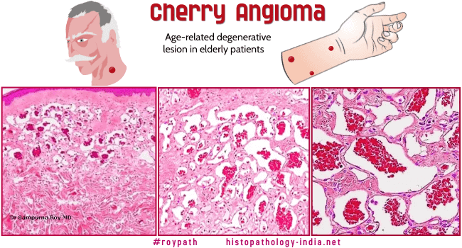
Cherry angioma is a common skin lesion usually noted in elderly adults. The lesion presents as single or multiple small, smooth, cherry red papules. This condition as commonly noted in elderly patients suggest that their occurrence is an age-related degenerative phenomenon. It is usually located on the trunk or upper extremities. Microscopic features: Small early lesion: Dilated interconnecting thin-walled vessels in the papillary dermis. Older lesion: Polypoid lesion; loss of rete ridges ; interconnecting vascular channels in the subepithelial connective tissue ; scant intervening stroma ; a collarette at the periphery.
|
|
|
Visit:- Infectious Disease Online

Consultant Histopathologist (Kolkata - India)
|
![]()
Copyright © 2022 histopathology-india.net

