

Custom Search
|

|
Infectious Disease Online Pathology of Varicella (Chickenpox) - Varicella-Zoster Virus
|
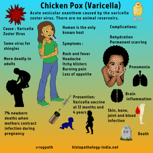
Varicella (chickenpox) is an acute vesicular exanthem caused by the varicella-zoster virus, an agent that has a worldwide distribution and for which humans are the only known host.There are no animal reservoirs.Varicella-zoster virus (HHV3) is a neurotrophic alpha-herpesvirus. Varicella-zoster virus (VZV) is named after "Varicella" , an alternative name for chickenpox (the primary Varicella- zoster virus infection) "Zoster", another name for shingles (reactivation of latent Varicella-zoster virus infection). Although all age groups are susceptible, in temperate zones chickenpox affects mostly children and in the tropics mostly young adults.The virus, which is spread through inhalation of droplets or by direct contact is highly contagious from about 24 hours before the initial eruption to a week of more thereafter.Although infection with varicella-zoster virus establishes lifelong immunity and chickenpox does not recur, the latent viral genome may be activated years later to cause shingles.Nonimmune persons (usually children) are susceptible to primary infection with varicella-zoster virus. Chickenpox begins as a "silent" infection of the nasopharynx, with local replication of varicella-zoster virus. After an incubation period of 10 to 23 days, the virus enters the bloodstream, causing viremia and a sudden onset of fever, malaise, and anorexia. In the circulation it seeds reticuloendothelial cells, an effect that leads to secondary waves of viremia. The virus then disseminates to skin and viscera, and within 24 hours a red maculopapular eruption develops, usually on the upper trunk and face. The papules rapidly become clear vesicles with an erythematous base. In the next 24 hours, the vesicles become cloudy, the eruption begins to itch, and scratching may rupture the vesicles. Separate crops appear for 3 to 6 days and spread peripherally. After the last crop, the scabs heal withour scarring. Though vesicles of the skin are generally painless, painful lesions may develop on mucous membranes, such as the cornea and tympanic membrane. Complications include pneumonia, encephalitis, hepatitis, carditis, keratitis, orchitis, arthritis, hemorrhages and acute encephalopathy with fat accumulation in the viscera (Reye’s syndrome). Histologically, the skin lesions initially show ballooning of epidermal cells. Later, unilocular vesicles containing proteinaceous fluid, degenerating cells and syncytial giant cells are seen. Cowdry type A intranuclear inclusions are seen in epidermal cells, endothelial cells of superficial capillaries, reticuloendothelial cells, and fibroblasts. The affected organs in chicken pox exhibit spherical foci of coagulative necrosis. At the margin of these necrotic foci, surviving cells contain intranuclear inclusions.
|
|
|

![]()
Copyright © 2022 histopathology-india.net