

Custom Search
|

|
Infectious Disease Online Pathology of Cryptosporidiosis
|
|
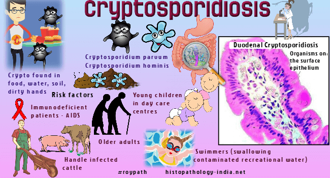
Cryptosporidiosis or "Crypto" is an
illness caused by Cryptosporidium
( a protozoan parasite) that
that can live in the
intestine of humans and animals and is passed in the stool of an infected
person or animal.
Cryptosporidium is an important cause of infectious diarrhea in humans and cattle. Cryptosporidium is most prevalent in AIDS patients but also has been found in children in day care centres, mental institution patients and international travellers. The majority of human infections are due to either Cryptosporidium hominis (C. hominis) and/or Cryptosporidium parvum (C. parvum). Mode of infection: It may be contracted through infected animals or from contaminated food and water. When the encysted Cryptosporidium are ingested, after exposure to acid in the stomach and digestive enzymes in the proximal duodenum, sporozoites are released from the exocyst. Sporozoites have a lectin on their surface that mediates adherance to intestinal epithelial cells. Malabsorption and diarrhoea occur when sporozoites disrupt the microvilli and enter the the cytoplasm of the epithelial cells. Cryptosporidium sporozoites also travel up the biliary tree to infect the epithelial cells that line the gallbladder and bile ducts. Acalculous cholecystitis and/or sclerosing cholangitis may then develop. Site: Gastrointestinal/hepatobiliary distribution of organisms: Small intestine, large intestine, pancreas, stomach, esophagus, extrahepatic and intrahepatic bile ducts. Jejunum is the favoured site. Clinical features: Transient watery diarrhea in normal children and debilitating diarrhea in patients with AIDS. Since some authors have demonstrated that Cryptosporidium is one of the most common causative microorganisms associated with sclerosing cholangitis in HIV patients, Cryptosporidium infection in the biliary tract should always be excluded in patients with HIV who have abnormal liver laboratory findings and cholangiography, even in the absence of chronic diarrhea. Investigations: The serologic tests and stool smears stained with acid fast are the conventional methods for detection of Cryptosporidium infection. However, morphologic diagnosis of Cryptosporidium by hematoxylin-eosin and electron microscopy has also been used. By electron microscopy, the characteristic morphologic findings of Cryptosporidium and its attachment to the luminal bile duct epithelium are diagnostic. Histological features: 2-5 micometer basophilic spherical structures (oocysts) adhere to the apical brush border of the absorptive epithelial cells of the intestine. Large numbers of oocysts are visible on the luminal surface of the epithelium lining the villi or in colonic crypts. The backround small bowel mucosa shows variable changes including blunting of villi and cryptitis. Special stains: These are well demonstrated by special stains (Giemsa, Gram stain, PAS and silver methenamine, but not acid fast stain). Differential diagnosis:
Unlike cryptosporidiosis in Cystoisosporiasis
the organism multiplies deep within the absorptive cells. |
Further reading: Pathogenesis of Cryptosporidium parvum infection.Host association of Cryptosporidium parvum populations infecting domestic ruminants in Spain. |
|
|

![]()
Copyright © 2022 histopathology-india.net