Custom Search

Dermpath-India

Pathology of Cutaneous Lymphadenoma

Dr Sampurna Roy MD      

Dermatopathology Quiz Case: 212   

Answer - Cutaneous Lymphadenoma


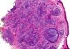
Cutaneous lymphadenoma is a rare tumour with distinctive histologic features.

This entity was originally described as lymphoepithelial tumour of the skin by Santa Cruz and Barr in 1987.

It was renamed cutaneous lymphadenoma in 1991.

Ackerman et al considered that cutaneous lymphadenoma is a 
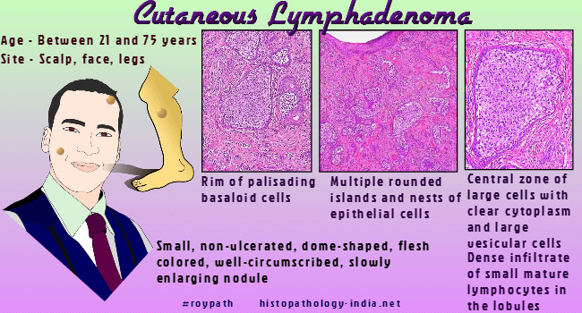
variant of trichoblastoma ("adamantinoid trichoblastoma").

Clinical presentation:
 A single, small, non-ulcerated, dome-shaped, flesh-colored,  well-circumscribed, slowly enlarging nodule.

The lesion has usually been present for many months or years.

Age and sex: Usually between 21 and 75 years without a sex predominance.

Site:  Face and scalp regions or legs.


Dermatopathology  Case: 151

Educational Gif – Pathology of Cutaneous Lymphadenoma

Microscopic features:

Multiple variably sized round to irregular islands or nests of epithelial cells ;

Tumour lobules consist of an outer rim of palisading basaloid cells surrounding a central zone of large cells with clear cytoplasm and large vesicular cells ;

Dense infiltrate of small, mature lymphocytes within the islands ;

Tumour lobules embedded in a loose-to-dense fibrous stroma ;  

Few scattered lymphocytes in the stroma ;

Focal follicular differentiation and sometimes sebaceous differentiation;

Focal mucinosis, osteoma formation or prominent sweat gland duct component in some cases ;

Central keratinization in some lobules.

Immunohistochemistry: 

Clear epithelial cells in the lobules and the basaloid peripheral cells: Cytokeratin positive.

Lymphoid component reacted with common leukocyte antigen (CLA) and  T-cell markers (UCHL-1, CD3).  

Some CD30+ large clear cells may be present. 

Many large epithelial cells in the lobules and some dendritic cells in the stroma : S-100 positive.

Differential diagnosis:

Clear cell syringoma ;  clear cell basal cell carcinoma ; lymphoepithelioma-like carcinoma of skin.

Cutaneous lymphadenoma does not tend to recur after surgical excision.

Dermatopathology Quiz Case


Further reading:

Cutaneous lymphadenoma.

Mohs micrographic surgery for cutaneous lymphadenoma.

Cutaneous lymphadenoma: a case report and immunohistochemical study.

A cutaneous adnexal neoplasm with features of adamantinoid trichoblastoma (lymphadenoma) in the benign component and lymphoepithelial-like carcinoma in the malignant component: a possible case of malignant transformation of a rare trichoblastoma variant.

Cutaneous lymphadenoma: a rare clinicopathological entity.

Cutaneous lymphadenoma.

Cutaneous lymphadenoma.

Lymphoepithelial tumor of the skin (abstr.). J Cutan Pathol 1987; 14: 345.


Visit:- Infectious Disease Online

Dr Sampurna Roy  MD

Consultant  Histopathologist (Kolkata - India)


Copyright © 2022  histopathology-india.net