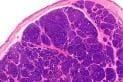
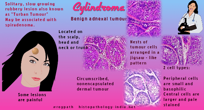
Cylindroma is a benign tumour in which
apocrine and trichoepitheliomatous differentiation has been noted
indicating complex hair follicle (folliculo-sebaceous-apocrine) rather
than eccrine differentiation.
Visit:
Cylindroma of the External Ear
Clinical presentation:
Clinically, the tumour usually presents as a solitary, slow growing
rubbery lesion.
The multiple form
is autosomal dominantly inherited on cyld1 gene on chromosome 16. This
form is also known as 'turban tumour' and may be associated with
spiradenoma. Some of these lesions are painful. This
condition is associated with trichoepitheliomas and milia.
Site:
Usually located on the
scalp, head and neck or trunk.
Microscopic features: Image
Link1
;
Image Link2
;
Image Link3 .
Histologically cylindroma is a circumscribed, non-encapsulated , dermal
tumour without attachment to the epidermis . The lesion is composed of
numerous oval and polygonal nests arranged in an interlocking 'jigsaw-like' pattern. There are two cell types, the peripheral cells are small
and basophilic and central cells are larger and pale stained. Small ductal lumina may be present. Thick PAS positive hyaline bands surround
tumour islands. Hyaline droplets within nests.
Malignant Cylindroma:
Cylindromas occasionally undergo malignant transformation. Malignant
cylindromas are characterised by islands of cells displaying marked
nuclear pleomorphism. There is increase in mitosis and many abnormal
forms are identified. The tumour shows invasion into the surrounding
tissue and loss of the delicate hyaline sheath.
Spiradenoma:
click ;
Spiradenocarcinoma:
click
Clinical image(NEJM):
Image Link
Image Links(Dr Weems):
Image1 ;
Image2
| Spiradenocylindrocarcinoma:
a malignant hybrid tumor.J Cutan Pathol. 2005 Jul;32(6):453
BACKGROUND: Eccrine
spiradenomas and cylindromas are benign, slowly growing, cutaneous adnexal
neoplasms, recently hypothesized to arise from a common pluripotential cell;
malignant variants are rare. We found 48 cases of malignant spiradenomas and
33 cases of malignant cylindromas reported in the English literature. Benign
tumors demonstrating both spiradenomatous and cylindromatous features have
been infrequently reported. Recent immunohistochemical studies of these two
tumors have provided compelling evidence that these two tumors may merely
represent a single tumor type with a spectrum of histological features. CASE
REPORT: We describe two cases of a malignant variant of this rare hybrid
tumor occurring in a 62-year-old male and a 72-year-old female. CONCLUSION:
We propose the term 'spiradenocylindrocarcinoma' to describe malignant
tumors with features of both a spiradenoma and a cylindroma. In conjunction
with histological features of malignancy, p53 and Ki-67 immunohistochemical
staining may provide helpful clues in determining the malignant potential of
this tumor. Further studies are needed to clarify the biologic behavior of
such lesions.
Fine needle aspiration cytology of breast
cylindroma in a woman with familial cylindromatosis: a case report.
BACKGROUND: That sweat gland type tumors occur
occasionally in the breast is not surprising, as the breast and
cutaneous sweat glands are embryologically related. Cylindromas
present most commonly as solitary and sporadic dermal nodules on the
face and scalp. Cases of multiple cylindromas are dominantly
inherited, and the neoplasms are referred to as "turban tumors" when
multiple lesions cover the scalp. Primary cylindroma of the breast has
been reported once in the past. To the best of our knowledge, the fine
needle aspiration cytology of primary breast cylindroma and its
occurrence in the setting of familial cylindromatosis have not
previously been reported. CASE: A 59-year-old woman presented with an
ill-defined left breast mass. She had a personal and family history of
dermal cylindromas on the head and face. Fine needle aspiration
cytology demonstrated small, uniform cells with oval nuclei and finely
granular cytoplasm, with some cells arranged around conspicuous
cylinders of dense, acellular material. Excisional biopsy was
recommended to exclude adenoid cystic carcinoma. Tissue biopsy
revealed a benign cylindroma arising in breast parenchyma. CONCLUSION:
Fine needle aspiration cytology of cylindroma very closely mimics that
of adenoid cystic carcinoma. Although extremely rare, primary breast
cylindroma is another entity to be included in the cytologic
differential diagnosis of bland, basaloid cells associated with
globular, extracellular material, a finding most commonly associated
with adenoid cystic carcinoma.
|
|