HISTOPATHOLOGY INDIA.COM

                        Adult Respiratory Distress Syndrome

    Dr  Sampurna Roy  MD


  Gastrointestinal Stromal Tumour


http://www.histopathology-india.net/Infection.htm


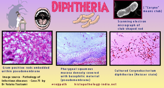
Diphtheria is an acute disease that results from a localized infection and a systemic toxemia caused by exotoxin -producing strains of Corynebacterium diphtheriae.

The name is derived from the Greek words koryne (club) and diphtheria (leather), the latter referring to the grey membrane at the site of infection.

Diphtheria is amenable to virtually complete eradication by routine immunization with diphtheria toxoid.

Even in medically advanced, however, diphtheria may occur when immunization procedures breakdown because of war or complacency.  An important epidemic occurred in Texus in 1970.

The low-grade endemicity of diphtheria depends on the occurrence of toxigenic Corynebacterium diphtheriae in the nasopharynges of a small number (fewer than 1%) of asymptomatic human carriers.

Transmission to nonimmune individuals usually occurs by the respiratory route.

In diphtheria, unlike food poisoning or tetanus, a parasitic relationship exists between the causative microorganism, C diphtheriae, and its host. 

Image1 Image2  ;  Image3  ;  Image4  ;  Image5  ;  Image6 (Dr Tsutsumi)

Clinical Presentation:

Diphtheria is a composite of a local inflammation and a systemic intoxication.

Toxin produced locally by toxogenic strains of C. diphtheriae is responsible for an inflammatory reaction on body surface at the site of infection (usually the oral pharynx, from which the process often extends to the nose or larynx).

Occasionally the tracheal, esophageal, or gastric mucosa is involved as well.

Less commonly, but particularly in the tropics, cutaneous trauma or burns may be the site of diphtheria.

The umbilical cord (in diphtheria neonatorum), the genital tract, and the conjuctivae are rare sites.

Unlike streptococcal tonsilitis, diphtheria is often insidious in onset and may be preceded by 2 to 3 days of listlessness, malaise, and headache before local symptoms occur.

Cervical adenopathy seems out of proportion to the pharyngeal lesion.

Soon small gray or white patches of exudates appear on the pharyngeal mucosa, usually over the tonsils.

These enlarge and coalesce and, with the accumulation of blood, become gray or black.

This exudate constitutes the characteristic diphtheritic membrane, which consists of leukocytes and numerous bacteria enmeshed in a dense network of fibrin.

The epithelial surface becomes necrotic and densely adherent to the overlying membrane ; this adherence explains why raw bleeding points are exposed when the membrane is forcibly removed.

If particularly extensive, the local process may produce mechanical respiratory obstruction, stridor, and even asphyxiation.

The local inflammatory process and its mechanical consequences are less important in the evolution of diphtheria than is the profound toxemia that characterizes the infection.

Diphtheria toxin, produced by C. diphtheriae in response to infection of that organism by a specific bacteriophage, is a potent inhibitor of cellular protein synthesis.

It is readily absorbed from the point of production into the blood stream, and its effects are noted in many organs and systems throughout the body.

Clinically apparent weakness or paralysis of limbs is rare. Neuropathic manifestations of diphtheria are usually temporary and disappear within 2 or 3 months if the patient survives.

Organs involved:

Lymph Node:  The lymphoid tissues both in regional lymph nodes and systemically (as in the spleen) undergo hyperplasia with the development of prominent germinal centers that are often centrally necrotic.

Heart:  Diphtheria toxin is particularly toxic to the myocardium.

In the early stages, interstitial edema, cloudy swelling of myocardial fibers, and the accumulation of fine cytoplasmic granules of lipid are seen microscopically.

Later these changes become widespread and are more severe.

Myocardial fibers eventually undergo necrosis, and a focal interstitial myocarditis with exudation of mononuclear cells occurs.

Cardiac involvement, either acutely in the form of cardiovascular collapse or as an arrhythmia or more chronically in the form of congestive heart failure, is the most common threat to life in diphtheria.

Kidney:  A nonspecific, nonsuppurative interstitial nephritis is frequent in diphtheria and is believed to be responsible for the proteinuria often observed. The renal lesion usually resolves completely in patients who recover.

Liver:  The liver is characteristically enlarged, hepatocytes exhibit cloudy swelling and less commonly focal necrosis.

Nerves:  Diphtherial toxin has a special affinity for peripheral nerves. Toxic effects are manifested in degeneration or even destruction of myelin sheaths. Axis cylinders undergo swelling and rarely necrosis. The paralytic effects of diphtheritic neuropathy are often sharply localized. Paralysis of the voluntary muscles of the palate may produce a peculiar nasal quality of the voice and a tendency to regurgitate fluids through the nose.

Hypopharynx:  Hypopharyngeal involvement may lead to aspiration pneumonia.

Eye:  Involvement of extraocular muscles may produce diplopia, and involvement of the ciliary body may result in defective visual accommodation.


Abstracts:

Emergence of an invasive clone of nontoxigenic Corynebacterium diphtheriae in the urban poor population of Vancouver, Canada.J Clin Microbiol. 2006 May;44(5):1625-9

Mapping and comprehensive analysis of the extracellular and cell surface proteome of the human pathogen Corynebacterium diphtheriae.
Proteomics. 2006 Apr;6(8):2465-76

Predictors of outcome in patients with diphtheria receiving intensive care.Indian Pediatr. 2006 Feb;43(2):155-60.

Infections with Corynebacterium diphtheriae: six years' experience at an inner London teaching hospital.Br J Biomed Sci. 2005;62(1):1-4.

Clinical features and predictors of diphtheritic cardiomyopathy in Vietnamese children.Clin Infect Dis. 2004 Dec 1;39(11):1591-8.  

Diphtheria: the patch remains.J Laryngol Otol. 2003 Oct;117(10):807-10.

Diphtheritic myocarditis: clinical and laboratory parameters of prognosis and fatal outcome.Ann Trop Paediatr. 2000 Sep;20(3):209-15

Atrioventricular block in a toxic child: do not forget diphtheria.Pediatr Cardiol. 2000 May-Jun;21(3):282-3

Nontoxigenic corynebacterium diphtheriae: an emerging pathogen in England and Wales?Emerg Infect Dis. 2000 Nov-Dec;6(6):640-5

Fulminant diphtheria myocarditis in an unvaccinated preschool child.Klin Padiatr. 1999 Nov-Dec;211(6):462-4

Respiratory diphtheria in three paediatric patients.Med J Malaysia. 1999 Sep;54(3):377-82

Diphtheroids and nontoxigenic Corynebacterium diphtheriae in the etiology of diphtheria.Mikrobiol Z. 1999 Jul-Aug;61(4):81-9

Diphtheria in the former Soviet Union: reemergence of a pandemic disease.Emerg Infect Dis. 1998 Oct-Dec;4(4):539-50.

Fatal respiratory tract diphtheria apparently caused by nontoxigenic strains of Corynebacterium diphtheriae.Eur J Clin Microbiol Infect Dis. 1997 Nov;16(11):816-20

The return of Corynebacterium diphtheriae: the rise of non-toxigenic strains.J Hosp Infect. 1995 Jun;30 Suppl:306-12

Resurgence of diphtheria.European Journal of Epidemiology.Volume 11, Number 1 / February, 1995

Infective endocarditis due to nontoxigenic Corynebacterium diphtheriae: report of seven cases and review.Clin Infect Dis. 1993 Feb;16(2):271-5

Diphtheria toxin entry into cells is facilitated by low pH.J Cell Biol. 1980 Dec;87(3 Pt 1):828-32.

Custom Search

Your Banner
Pathopedia-India.com:

Contents ; Introduction of Pathology ; An outline of Diagnostic Techniques available in Pathology ; Cellular Injury ; Diagram showing Structural Changes in Reversible and Irreversible Cell Injury ; Autolysis; Heterolysis ; Necrosis; Coagulation (Coagulative) necrosis ; Caseative (Caseous) necrosis ; Liquefaction necrosis ; Fat necrosis ; Fibrinoid necrosis ; Apoptosis ; Gangrene ; Hyaline Change ; Atrophy ; Hypertrophy ; Hyperplasia ; Metaplasia ; Aplasia ; Hypoplasia ;Cellular Accumulations ; Accumulation of Glycogen, complex lipids and carbohydrates ; Pigments ; Melanin ; Pigments derived from Hemoproteins; Hemosiderin and Hemosiderosis ; Primary Hemochromatosis ; Hematin; Bilirubin; Lipofuscin; Mineral Dusts ; Silica ; Urate ; Amyloid ; Inflammation ; Inflammatory cells in acute and chronic inflammation ; Acute Inflammation; Types of Acute Inflammation; Chemical Mediators ; Chronic Inflammation; Wound Healing ; Circulatory Anatomy, Physiology and Regulation; Normal Fluid Balance; Edema; Morphology of Edema; Diagram showing Capillary System and Mechanisms of Edema Formation; Hyperemia and Congestion; Hemostasis and Thrombosis; Embolism; Fat Embolism; Air Embolism ; Decompression Sickness ; Amniotic Fluid Embolism ; Diagram showing Sources of Arterial Emboli ; Diagram showing Sources of Venous Emboli ; Infarction ; Diagram showing common sites of Systemic Infarction  from Arterial Emboli; Shock; Pathology of Shock; Diagram showing Complications of Shock; Hemorrhage;

September 2008
Pathopedia-India.com

Surgical-Pathology.com

Histopathology-India.net

Pathology-India.com

Pancreatic Pathology Online

Gall Bladder Pathology Online

Paediatric Pathology Online

Paraganglioma-Online

Endocrine Pathology Online

Eye Pathology Online

Ear Pathology Online

Cardiac Path Online

Lung Tumour-Online

Mesothelioma-Online

Pulmonary Pathology Online

Nutritional Pathology Online

Environmental Pathology Online

Pathology Quiz Online

Dermpath-India

GI Path Online

Soft Tissue Pathology

Case Index

Infectious Disease Online; INDEX: A-D ; INDEX: E-L ; INDEX: M-P INDEX: Q-Z ; FUNGAL DISEASE ; VIRAL DISEASE.

E-book - History of  Medicine with special reference to India

Actinomycosis

Cutaneous lesion associated  with AIDS

Amoebiasis (Entamoeba histolytica)

Anthrax Infection

Avian Influenza (Bird Flu)

Bacillary angiomatosis

Bartonellosis

Blackwater Fever

Blastomycosis

Botulism

Bronchopneumonia

Brucellosis

Buruli Ulcer

Campylobacter Infection

Capillariasis

Carbuncle

Cat Scratch Disease

Candidosis(Candidiasis)

Cestodes(Tapeworms)

Chagas' Disease

Chikungunya

Chlamydial Infection of the Genital Tract

Cholera

Chromomycosis

Clonorchiasis

Clostridium Perfringens Food Poisoning

Clostridial myonecrosis

Coccidioidomycosis

Cryptococcosis

Cryptosporidium

Cytomegalovirus infection

Dengue

Dermatophytosis

Eastern equine encephalitis

Ebola Virus Infection

Echinococcosis(Hydatid Disease)

Echovirus Infection

Enterobiasis

Epidemic Typhus

Epstein-Barr Virus infection

Epstein-Barr Virus  Related Malignant Tumours

Erythema chronicum migrans

Escherichia coli  Infection

Filariasis

Flavivirus

Giardiasis

Herpes Simplex Virus Infection

Histoplasmosis

Hookworm Infection

Human Papilloma Virus Associated Epidermal Lesions

Infective Endocarditis

Isosporiasis

Leishmaniasis

Listeriosis

Lobomycosis

Malaria

Meningococcal  Infection

Measles

Molluscum  Contagiosum

Mycobacterium Avium Intracellulare

Onchocerciasis

Pneumocystis Pneumonia

Protothecosis

Pseudomembranous Colitis (Clostridium difficile)

Pulmonary Infection

Rat Bite Fever

Rhinosporidiosis

Salmonellosis (Gastroenteritis and Septicemia)

Schistosomiasis

Shigellosis

Skin infections- (Histo-pathological patterns)