

Custom Search
|

|
Infectious Disease Online Pathology of Dracunculiasis (Guinea Worm Disease)
|
|
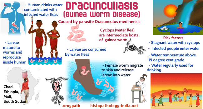
Syn: Guinea worm infection; Medina worm infection.
Dracunculiasis is caused by the parasite Dracunculus medinensis, or the guinea worm. The larvae are taken up by a freshwater crustacean of the genus cyclops (water fleas). They mature within the crustacean into elongated larvae infective for humans. These larvae are liberated by the gastric digestive juices when a person drinks water contaminated with parasite infected cyclops (water fleas). The larvae migrate through the wall of stomach or small intestine into the connective tissue of the abdominal wall. The male worm is small and probably dies after copulation. The female worm, which averages 1m in length, wanders out to the subcutaneous tissue, specially that of the feet and legs. A small papule appears, which later becomes a vesicle with a small ulceration in the center through which the embryo are liberated. The latter are usually present in milky fluid contained in a segment of the worm’s uterus that is extruded and breaks away from the body of the parasite. On rare occasions the worms may be found in joints and serosal cavities. The signs and symptoms of dracunculiasis are limited to the period of liberation of embryos. Pruritus and moderate edema may be accompanied by low-grade fever and urticarial rash, caused by an anaphylactic reaction to the products of the worm. If the worm dies or if the embryos are liberated into the tissues, a severe inflammatory reaction with abscess formation ensues. Eighteen of 20 countries were known in 1986 to have endemic dracunculiasis, i.e., Benin, Burkina Faso, Cameroon, Chad, Cote d'Ivoire, Ethiopia, Ghana, India, Kenya, Mali, Mauritania, Niger, Nigeria, Pakistan, Senegal, Sudan, Togo, and Uganda. Image Link Transmission of the disease in Yemen was documented in 1995, and the World Health Organization (WHO) declared Central African Republic endemic in 1995. As of the end of 2004, a total of 16026 cases of dracunculiasis were reported from 12 endemic countries (91% of these cases were reported from Ghana and Sudan, combined). WHO has certified 168 countries free of dracunculiasis, including Pakistan (1996), India (2000),Senegal and Yemen (2004). Asia is now free of dracunculiasis. The total of 27 cases in 2020 were reported from four countries: Angola (1 case), Chad (12 cases), Ethiopia (11 cases), Mali (1 case), South Sudan (1 case) and Cameroon (1 case, likely imported from Chad).
|
|
|

![]()
Copyright © 2022 histopathology-india.net