Gastrointestinal Stromal Tumour


http://www.histopathology-india.net/SoftTissuePath.htm

                       HISTOPATHOLOGY INDIA.COM           

               Path Quiz Case-33

      Diagnosis:  Atypical Fibroxanthoma

            Dr Sampurna Roy  MD

 
 January 2008
Surgical-Pathology.com

Histopathology-India.net

Pathology-India.com

Pancreatic Pathology Online

Gall Bladder Pathology Online

Paediatric Pathology Online

Paraganglioma-Online

Endocrine Pathology Online

Eye Pathology Online

Ear Pathology Online

Cardiac Path Online

Lung Tumour-Online

Mesothelioma-Online

Pulmonary Pathology Online

Nutritional Pathology Online

Environmental Pathology Online

Pathology Quiz Online

Dermpath-India

GI Path Online

Soft Tissue Pathology

Case Index

Infectious Disease Online; INDEX: A-D ; INDEX: E-L ; INDEX: M-P INDEX: Q-Z ; FUNGAL DISEASE ; VIRAL DISEASE.

Nodular fasciitis

Proliferative fasciitis

Proliferative myositis

Ischaemic fasciitis

Elastofibroma

Fibrous Hamartoma of Infancy

Infantile Myofibromatosis/ Myofibroma

Juvenile hyaline fibromatosis

Inclusion  Body Fibromatosis

Calcifying aponeurotic fibroma

Fibromatosis colli

Fibroma of tendon sheath

Desmoplastic fibroblastoma  

Storiform Collagenoma (sclerotic fibroma)

Giant Cell Collagenoma

Pleomorphic Fibroma

Angiomyofibroblastoma

Dermatomyofibroma

Cellular Angiofibroma

Giant Cell Angiofibroma

Fibromatosis

Lipofibromatosis

Solitary fibrous tumour

Inflammatory myofibroblastic tumour

Low grade myofibroblastic sarcoma

Myxoinflammatory fibroblastic sarcoma


               Case 33:Case history and images:


Elastofibroma is a benign slowly progressive reactive lesion involving abnormal elastogenesis.

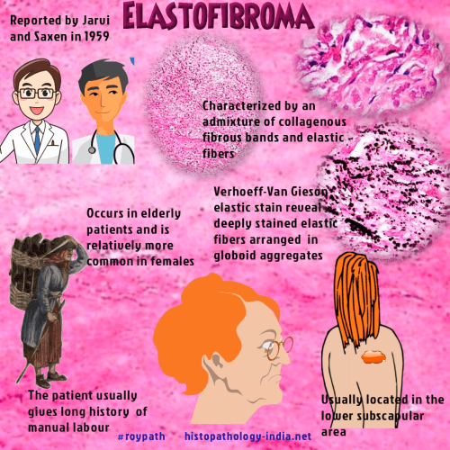
It was first reported by Jarvi and Saxen in 1959 and published in 1961.

The lesion usually occurs in elderly patients and is relatively more common in females. Most cases are unilateral.

Bilateral involvement is present in about 10% of all cases.

The patient usually gives long history
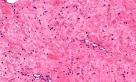
 of manual labour.

Elastofibroma is usually located in the lower subscapular area and is probably caused by friction between the inferior edge of the scapula and the underlying chest wall.

Cases have also been reported  in the deltoid muscle, hip, thigh and stomach.

Histologically, the lesion is characterized by an admixture of collagenous fibrous bands and elastic fibres. Elastin stains reveal branched or unbranched fibres with irregular serrated margins or fibres arranged in globoid aggregates. 

Electron microscopic studies revealed that the fibres consisted of granular aggregations of electron dense material along a central core surrounded by an amorphous matrix containing microfibrils. The collagen deposited in this disease is a mixture of Type I, II and III.

According to some authors it has been assumed there is an underlying genetic disposition or inherent enzymatic defect.

This benign reactive lesion has no tendency to recur and is usually treated by surgical excision.

Image Link1 ; Image Link2 ; Image Link3 ; Image Link4


Further reading:

Elastofibroma Dorsi- Acta Pathol Microbiol Scand 51:83, 1961

Elastofibroma. A correlated light and electron microscopic study. Virchows arch (Pathol Anat) 369 (2):127-140, 1982 

Elastofibromatous lesion of the stomach in a patient with elastofibroma dorsi.Am J Surg Pathol 9(3) : 233-237, 1985

Elastofibroma in Okinawa.A clinicopathologic study of 170 cases. Cancer 50(9):1794-1805,1982 

Elastofibroma. Disturbed elastic fibrillogenesis by periosteal-derived cells ? An immunoelectron microscopic and in-situ hybridization study. Hum Pathol 22: 1017-1029, 1991

"Aberrant elastic" in elastofibroma. An immunohistochemical and ultrastructural study.  Ultrastruct pathol 19: 45-50, 1995 

Elastofibroma of the stomach. Pathol Res Pract. 199 (9) : 637-639, 2003

Elastofibroma associated with high grade leiomyosarcoma of the soft tissue ;  a light and ultrastructural study of one case . J  Submicrosc Cytol  Pathol .35 (1) : 43-8,  2003


Soft Tissue Pathology;

Myxoid Tumours of Soft Tissue Classification of Soft Tissue Tumour;  Gross examination of soft tissue specimen ;  A practical approach to histopathological reporting of soft tissue tumours Grading of soft tissue tumours ; Lipomatous tumours ;Neural tumours ; Myogenic tumours ;Vascular tumours ;Fibroblastic/Myofibroblastic tumours ; Myofibroblastic tumours ;  Fibrohistiocytic tumours ; ChondroOsseous tumours ; Soft TissueTumours of Uncertain Differentiation ; Notochordal Tumour -Chordoma ;Extra-adrenal Paraganglioma ; Gastrointestinal Stromal Tumour ;

Your Banner
PULMONARY PATHOLOGY:

Congenital Cystic Adenomatoid  Malformation ; Acute Respiratory Distress Syndrome  ;Sarcoidosis ;Bronchiolitis ; Emphysema ; Bronchial Asthma ;Chronic Bronchitis Pulmonary Alveolar Proteinosis ; Lipid Pneumonia ; Pulmonary Hypertension ;Pulmonary edema ;Pulmonary Infection ; Pneumococcal Pneumonia ; Haemophilus influenza Infection;Klebsiella Pneumoniae ; Mycoplasma Pneumonia ; Pneumocystis Pneumonia ; Legionellosis ; Localized Fibrous Tumour of the Pleura ; Biphasic Epithelial/Mesenchymal Lung Tumours ; Pulmonary Carcinosarcoma ;Pulmonary Blastoma ; Large Cell Neuroendocrine tumour;