

Custom Search
|

|
Infectious Disease Online Pathology of Enterobiasis
|
|
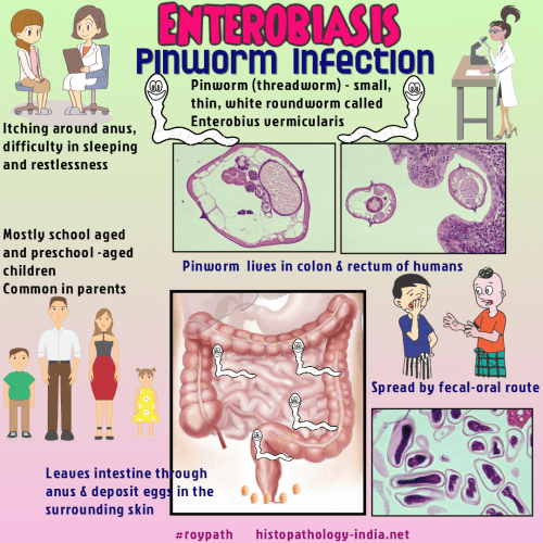
Syn: Pinworm ; Threadworm Enterobiasis is a very common parasitic disease of cosmopolitan distribution. The causative nematode is Enterobius vermicularis, formerly known as Oxyuris vermicularis, or commonly as the pinworm or threadworm.
The disease is acquired by ingestion of fully embryonated ova deposited by the female worm about the anus and transferred to other hosts by fecal contamination or to the same host to produce reinfection. The ova are very resistant to destruction, and the infection occasionally affects all persons in the same household. The life span of the worm is short (1.5 to 2 months), and the main problem is reinfection. The larvae are hatched from the fully embryonated ova on the region of the duodenum and then pass downward while they molt twice and mature, until they reach the ileocecal area. The adult worms attach themselves to the superficial portion of the mucosa of the terminal ileum, caecum, and appendix by their anterior end. The pathologic changes produced by the adult worms in this area are minimal, and their role in the pathogenesis of appendicular symptoms in some cases is not clear. The general impression is that, even when adult worms are present in acute appendicitis, they are not a significant cause of inflammation. The most important symptoms of the disease is intense pruritus ani caused by the migration of the adult female worm to the perianal region, when she lays her eggs. The adult male worm is unimportant in the pathogenesis, since it dies after copulation. Rarely, Enterobius vermicularis can be seen in extra- intestinal sites. In female the nematode migrates up the uterus and fallopian tubes to produce a foreign-body type of granulomatous inflammation most commonly seen in the surface of the ovaries, omentum, and peritoneum. This parasite apparently cannot survive outside its normal habitat, and the granulomatous reaction described occurs after death of the worms. Other less common ectopic sites include the urinary bladder, lung, and liver.
An association has been reported between enterobiasis and lower urinary tract infections, with the nematode acting as a carrier of enteric bacteria when it migrates from the perianal region to the urinary bladder. Diagnosis is made by identification of the ova by a cellophane-tape test.
|
|
|

![]()
Copyright © 2022 histopathology-india.net