HISTOPATHOLOGY INDIA.COM

               Path Quiz Case- 86

           Myxoid Tumours of Soft Tissue

  Dr  Sampurna Roy  MD

         Case history and images:


      DermPath-India

        Site created by

  Dr Sampurna Roy MD


http://www.histopathology-india.net/dermpath.htm


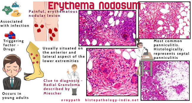
Erythema nodosum is a disease characterized by painful , erythematous nodular lesions. It is the most common panniculitis and histologically represents the prototype of a septal panniculitis.

Site:  Situated on the anterior and lateral aspect of the lower extremities and rarely on the arms, trunk, and calves, and on the neck and face.

Age:  Usually occurs in young adults. Cases have been reported in children and in elderly patients.

Pathogenesis:  It is not known and may be due to allergic response to infection or systemic disease.

Erythema nodosum is associated with the following conditions:

Infection : Streptococcal ;Meningococccal ;Campylobacter ; Salmonella ; Histoplasma ; Mycobacterial ; Dermatophyte; Yernia.

Inflammatory bowel disease ; Sarcoidosis ; Behcet disease ; Sweet's disease ; Carcinoma ; lymphoma ; leukemia.

Other triggering factors include drugs (Eg.  Sulphonamides ; penicillin ; oral contraceptive) and radiotherapy.

Microscopic features:  Image Link1 ; Image Link2 ; Image Link3 ; Image Link4 .

Septal panniculitis ;

Small foci of inflammatory cells extend into adjacent lobular fat ; 

Sometimes spillover of cells is prominent and include foam cells ;

There may be focal fat necrosis adjacent to the septa;

Septal infiltrate is composed of lymphoid cells, variable numbers of foreign body giant cells and occasional eosinophils and histiocytes ;

In fully developed lesions granulomas are present in the center of lobules ;

A  clue helpful to diagnosis of an early lesion of erythema nodosum is the finding of "radial" granulomas, first described by Miescher and consisting of clusters of neutrophils in collections of epithelioid histiocytes;

A few neutrophils  are present in the early lesion ;

Fibrous septa in the subcutaneous fat are widened by edema ;

Mild to moderate perivascular infiltrate of lymphocytes around vascular plexuses of the reticular dermis ;

Sometimes a definite vasculitis is present (lymphocytic or leukocytoclastic type).

 In late lesions septa widened by marked fibrosis ;  Fat lobules are  markedly reduced in size by widened fibrotic septa.

                 
Abstracts:

Histopathologic spectrum of erythema nodosum.: J Cutan Pathol. 2006 Jan;33(1):18-26.

Erythema nodosum.Dermatol Online J. 2002 Jun;8(1):4.

Panniculitis. Part I. Mostly septal panniculitis.J Am Acad Dermatol. 2001 ; 45(2):163-83; quiz 184-6.

Histopathologic features of erythema nodosum--like lesions in Behcet disease: a comparison with erythema nodosum focusing on the role of vasculitis.Am J Dermatopathol. 2000;22(5):379-90.

Erythema nodosum versus nodular vasculitis.Int J Dermatol. 1993 ;32(2) :108-12.

Miescher's radial granuloma. A characteristic marker of erythema nodosum. Am J Dermatopathol. 1989;11(5):434-42.


Your Banner

March 2008
Surgical-Pathology.com

Histopathology-India.net

Pathology-India.com

Pancreatic Pathology Online

Gall Bladder Pathology Online

Paediatric Pathology Online

Paraganglioma-Online

Endocrine Pathology Online

Eye Pathology Online

Ear Pathology Online

Cardiac Path Online

Lung Tumour-Online

Mesothelioma-Online

Pulmonary Pathology Online

Nutritional Pathology Online

Environmental Pathology Online

Soft Tissue Pathology

Case Index

Pathology Quiz Online

Dermpath-India

GI Path Online

Infectious Disease Online;

INDEX: A-D ; INDEX: E-L ; INDEX: M-P INDEX: Q-Z ; FUNGAL DISEASE ; VIRAL DISEASE.

E-book - History of  Medicine with special reference to India

 
Granulomatous Reaction Pattern of the Skin

Granuloma Annulare

Necrobiosis Lipoidica

Necrobiotic Xanthogranuloma   

Rheumatoid Nodule

Lupus Vulgaris

Cutaneous Sarcoidosis

Melkersson Rosenthal Syndrome

Annular Elastolytic Giant Cell Granuloma

Skin lesion in Crohn's Disease

Blastomycosis-like pyoderma

Foreign body granuloma

Rosacea

Interstitial Granulomatous Dermatitis

Interstitial Granulomatous Drug Reaction

Granulomatous T-cell lymphoma