Custom Search

Dermpath-India

Pathology of Hailey-Hailey Disease

Dr Sampurna Roy MD                               2022


Dermatopathology Quiz Case: 241

Answer - Hailey-Hailey Disease


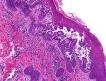
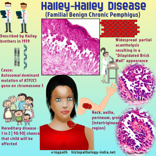
Hailey-Hailey disease (familial benign chronic pemphigus) is an autosomal dominant disorder with recurrent eruption of vesicles and bullae. 

It was originally described by the Hailey brothers in 1939.

The lesion is caused by mutations in the ATP2C1 gene encoding a novel Ca(2+) pump. 

The blistering dermatitis is characterized by acantholytic cells present in foci throughout at least half of the thickness of the epidermis.

Site:  

Predominantly involve the neck, groin, inframammary, perianal and axillary regions. 

Rarely oral, ocular, esophageal and vaginal cases have been reported.

Age:

Usually occurs in the second or third decade of life.

Clinical presentation:  

Usually presents as well demarcated plaques. Rare variants: Papular, annular, verrucous, and vesiculopustular variants.

Course:

This chronic disease is characterized by spontaneous remission with subsequent exacerbation.

Histopathological features: 

Histologically, Hailey-Hailey Disease has a characteristic 'dilapidated brick wall appearance'. [Elongated papillae covered by a layer of keratinocytes (villi) protruding into the bulla] .

Early:

Superficial perivascular infiltrate of mainly lymphocytes ; 

Changes are located in focal areas  in the lower half of the epidermis ;

Vacuoles are present between basal cells and spinous cells and between spinous cells.

Fully developed:

Moderately dense perivascular and interstitial infiltrate of lymphocytes ; 

Edema of the papillary dermis ; Acanthosis ; Suprabasal blister ; 

Acantholytic dyskeratotic cells, some with polygonal outlines, above the basal layer either singly or in clumps lining the clefts  ;  

Present throughout at least the lower half of the epidermis ;  

Infundibular epidermis and epithelial structures of adnexa spared by the acantholytic process ; 

Scale-crusts ;  

Neutrophils are sometimes numerous within the vesicle or in the surface crust.

Late:

All the features of fully developed lesions with the addition of a mixed-cell infiltrate, acantholytic cells increasingly dyskeratotic, and more prominent scale-crusts.


Differential diagnosis:

Hailey-Hailey variant of Grover's disease:

Narrow vesicle involving not more than a few rete ridges. Hailey-Hailey disease is more florid.

Example: More confluence, more markedly thickened epidermis, more acantholytic cells, more dyskeratotic cells with polygonal outlines, more vesiculation, and more scale-crusts.

Pemphigus vulgaris:  

Less acantholysis and acantholytic cells are confined to the suprabasal region;

Some cells show more prominent dyskeratosis ;

Epidermis of relatively normal thickness ; 

Infundibular epidermis & epithelial structures of adnexa are involved by the process;

There are no scale-crusts.

Dermatopathology Quiz Case


Further reading

What's in a name?: Hailey-Hailey disease.

Type 2 segmental Hailey-Hailey disease with systematized bilateral arrangement.

Efficacy of the melanocortin analogue Nle4-D-Phe7-α-melanocyte-stimulating hormone in the treatment of patients with Hailey-Hailey disease.

Incidental cutaneous reaction patterns: epidermolytic hyperkeratosis, acantholytic dyskeratosis, and hailey-hailey-like acantholysis: a potential marker of premalignant skin change.

Novel Mutation in ATP2C1 Gene in a Japanese Patient with Hailey-Hailey Disease.

Keratinocytes cultured from patients with Hailey-Hailey disease and Darier disease display distinct patterns of calcium regulation.

Severe impairment of quality of life in Hailey-Hailey disease.

Genital benign chronic pemphigus (Hailey-Hailey disease) presenting as condylomas.

Hailey-Hailey disease: a widespread abnormality of cell adhesion.

Histologic findings of Hailey-Hailey disease in a patient with bullous pemphigoid.  


Visit:- Infectious Disease Online

Dr Sampurna Roy  MD

Consultant  Histopathologist (Kolkata - India)


Copyright © 2002-2022  histopathology-india.net