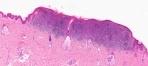
DermPath-India

     Site created by

 Dr Sampurna Roy MD


http://www.histopathology-india.net/dermpath.htm

                               HISTOPATHOLOGY INDIA.COM

                  Desmoplastic/Spindle cell
/Neurotropic Melanoma

       Dr. Sampurna Roy  MD

 
Web www.histopathology-india.net
 
  Gastrointestinal Stromal Tumour


http://www.histopathology-india.MelanocyticTumours.htm

 May 2007
Surgical-Pathology.com

Histopathology-India.net  

Eye Pathology Online

Cardiac Path Online;

Pulmonary Pathology Online

Pathology Quiz Online;

Dermpath-India;

GI Path Online

Mesothelioma-Online;

Soft Tissue Pathology;

Infectious Disease Online;

INDEX: A-D ; INDEX: E-L ;INDEX: M-P ;INDEX: Q-Z ; FUNGAL DISEASE ; VIRAL DISEASE.

Melanocytic tumours
              
1. Acquired Melanocytic Naevus

Ancient Naevus  
Halo naevus
Balloon cell naevus
Combined Naevus
Recurrent melanocytic naevus

                   
2. Spindle and epithelioid cell naevi

Spitz naevus
Pigmented spindle cell naevus

3. Blue naevi

Common blue naevus 
Cellular blue naevus


4. Dermal melanocytoses

Naevus of Ota

Naevus of Ito  
Mongolian spot  

5. Congenital melanocytic naevi

6. Dysplastic melanocytic naevi                                           

       
The lesion is characterized  by amelanotic ring  upto several milimeters in width around a melanocytic naevus. Image Link(DermAtlas):click

(Clinical D/D: Meyerson's Naevus -eczematous halo around the naevus).

Histologically, a dense lymphocytic infiltrate is present in the dermis which may obscure the melanocytic component.

  click on the image

Nevus cells are present in nests or singly within the lymphocytic infiltrate. The number of nevus cells depend on the stage at which the biopsy was taken.

Some cells show cytological atypia. (D/D: In melanoma such a dense diffuse lymphocytic infiltrate is usually not present.)

Some macrophages and factor XIIa-positive dendrocytes may be present in the infiltrate.

In some cases, the  melanocytes may be epithelioid in type and may display some cytological atypia. The lymphocytes are mostly CD8- positive lymphocytes. These cells express CD69 and tumour necrosis factor -alpha.

Depigmented halo shows an absence of melanin pigment and melanocytes in the basal layer.

(Both humoral and cell mediated immunity are involved in the rejection of nevus cells and formation of halo naevus.)

             Image Links1(Dr Weem)  Image2  

Halo nevus or halo phenomenon? A study of 142 cases.J Cutan Pathol. 1995;22(4):342-8.


Pigmented melanocytic lesions causing diagnostic problems

Prognostic parameters of melanoma

Lentigo maligna melanoma

Superficial spreading melanoma

Nodular melanoma

Acral lentiginous melanoma

Desmoplastic /Spindle cell /
Neurotropic melanoma

Naevoid melanoma

Balloon cell melanoma