Custom Search


Dermpath-India

Pathology of Hidradenoma

Dr Sampurna Roy MD                    2022


Dermatopathology Quiz Case: 248

Answer -  Hidradenoma


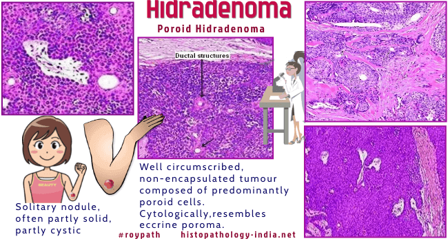
Syn: Clear cell hidradenoma,  
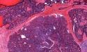
solid and cystic hidradenoma or dermal acrospiromas.

Hidradenoma is a benign dermal appendage tumour.

Hidradenomas may be of eccrine or apocrine origin.

Solitary, nodule often partly solid, partly cystic.

Site:  

Located in any part of the body. There is a slight predilection for the head, face, and upper extremities.


Microscopic features:  

Well-circumscribed dermal tumor with a grenz zone between the tumour and the epidermis ;

Variously shaped tumour islands in the dermis ;

May extend into the subcutis ; 

Combination of solid and cystic areas ;

Cystic spaces form as a result of tumour cell degeneration ;

The intervening stroma varies from delicate vascularized  fibrous tissue to dense hyalinized collagen ;  

Minimal epidermal connection may be present ; 

Foci of squamous differentiation; 

Ductal differentiation and intracytoplasmic lumina ; 

Mitotic figures are often present which is of little prognostic significance.

Two types of cells -  One cell type is polyhedral with a rounded nucleus and slightly basophilic cytoplasm. The second cell type is generally round with clear cytoplasm.

Variants:  Clear cell ; Poroid ; Oncocytic ; Epidermoid, and Pigmented. 

Clear cell hidradenomas are characterized by prominent clear cells which is present in less than one third of hidradenomas. Mucus producing cells are found in some cases.  The clear cells contain glycogen and periodic acid-Schiff-positive, diastase-resistant material, but no lipid.

Poroid hidradenomas are well circumscribed , non-encapsulated tumours composed of predominantly poroid cells. 

Cytologically, this variant resembles an eccrine poroma.

Atypical hidradenomas - cytological atypia or infiltrative margins; higher rate of recurrence and malignancy. The significance of atypical nuclear changes in benign eccrine acrospiromas: a clinical and pathological study of 18 cases.


Differential diagnosis:

It is sometimes difficult to distinguish solid cellular hidradenomas without duct-like structures and glomus tumour without a vascular pattern.

Clear cell hidradenoma:    Differential Diagnosis- Clear cell tumours of skin ; Hidradenocarcinoma. (Clear cell hidradenoma: a mimic of metastatic clear cell tumors).  

Nodular hidradenoma can recur after inadequate excision.


Further  reading:

Clear cell hidradenoma

Clear cell hidradenoma: An unusual tumor of the oral cavity

Clear Cell Hidradenoma of the Axilla: a Case Report with Literature Review

Malignant Nodular Hidradenoma of the Eyelid: A Rare Sweat  Gland Tumor

Nodular hidradenoma of male breast: Cytohistological correlation

Giant benign nodular hidradenoma of the shoulder: A rare tumor in orthopedic practice

Tubulopapillary hidradenoma: A rare case with cyto histopathological correlation

Clear cell hidradenoma: a mimic of metastatic clear cell tumors.

Poroid hidradenoma.

Poroid hidradenoma. Report of a case with cytologic findings on fine needle aspiration.

Poroid hidradenoma. A case presentation.


Visit:- Infectious Disease Online

Dr Sampurna Roy  MD

Consultant  Histopathologist (Kolkata - India)


Copyright © 2002-2022  histopathology-india.net