Gastrointestinal Stromal Tumour


http://www.histopathology-india.net/SoftTissuePath.htm

                                        HISTOPATHOLOGY INDIA.COM

                 Fibrous Hamartoma of Infancy

            Dr Sampurna Roy MD

 
 August 2009
Histopathology-India.net

diagnostic histopathology. blogspot.com

Pathopedia-India.com

Surgical-Pathology.com

Pathology-India.com

Pancreatic Pathology Online

Gall Bladder Pathology Online

Paediatric Pathology Online

Paraganglioma-Online

Endocrine Pathology Online

Eye Pathology Online

Ear Pathology Online

Cardiac Path Online

Lung Tumour-Online

Mesothelioma-Online

Pulmonary Pathology Online

Nutritional Pathology Online

Environmental Pathology Online

Pathology Quiz Online

Dermpath-India

GI Path Online

Soft Tissue Pathology

Case Index

Infectious Disease Online; INDEX: A-D ; INDEX: E-L ; INDEX: M-P INDEX: Q-Z ; FUNGAL DISEASE ; VIRAL DISEASE.

E-book - History of  Medicine with special reference to India.

Basic Pathology Blog

Myxoid Tumours of Soft Tissue

Classification of Soft Tissue Tumour

Gross examination of soft tissue specimen          

A practical approach to histopathological reporting of soft tissue tumours

Grading of soft tissue tumours

Lipomatous tumours

Neural tumours

Myogenic tumours

Fibroblastic/ Myofibroblastic tumours

Myofibroblastic tumours

Fibrohistiocytic tumours

ChondroOsseous tumours

Soft TissueTumours of Uncertain Differentiation               

Notochordal Tumour - Chordoma

Extra-adrenal Paraganglioma

Gastrointestinal Stromal Tumour

Nodular fasciitis

Proliferative fasciitis

Proliferative myositis

Ischaemic fasciitis

Elastofibroma

Fibrous Hamartoma of Infancy

Infantile Myofibromatosis

Juvenile hyaline fibromatosis

Calcifying aponeurotic fibroma

Fibromatosis colli

Fibroma of tendon sheath

Desmoplastic fibroblastoma

Storiform Collagenoma 

Giant Cell Collagenoma 

Angiomyofibroblastoma

Dermatomyofibroma

Cellular Angiofibroma

Giant Cell Angiofibroma

Fibromatosis

Lipofibromatosis

Solitary fibrous tumour

Inflammatory myofibroblastic tumour


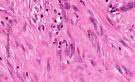
Inclusion body fibromatosis (Infantile digital fibromatosis) is a rare, benign fibrous growth of childhood. It was identified as a distinct entity by Reye in 1965.

This lesion has also been referred to as Reye tumor, infantile digital fibroma and recurrent digital fibrous tumour of childhood.

Age:  These lesions are usually present in young infants of either sex. Most of the cases are diagnosed within the first year of life and up to one third of cases are congenital. Some cases have been reported in adults (non-digital sites).

Site:   These lesions usually present as single or multiple nodules involving the extensor surfaces, most commonly the dorsal and lateral aspect of the distal or middle phalanx. Thumbs and great toes are usually spared.                               Cases have been reported in extradigital locations such as tongue, breast, arm and leg.

Gross features:  Pale, hard , poorly circumscribed, nontender nodule with a firm broad-base. The lesion is covered by intact skin and is often less than 1cm in diameter.

Microscopic features

The lesion replaces most of the dermis.
It is a non-encapsulated tumour composed of moderately cellular whorls, sheets and interlacing bundles of eosinophilic myofibroblasts set in a collagenous backround.

The most characteristic feature is the presence of 'cytoplasmic round eosinophilic inclusions'  in a paranuclear position. The inclusions are sometimes surrounded by a clear halo. These are slightly larger than red blood cells. With maturation of the lesion there may be decrease in number of inclusions.  These inclusions are composed of actin which has been confirmed by immuno-electron microscopy.

Adnexal structures and fat may be entrapped in the fibrous proliferation. The lesion may extend to the periosteum and erode it. There is no invasion of bone.

[The following features may be indicative of tumour recurrence:    Incomplete surgical or histological resection; Patient's age more than 5 years ; Mitotic index of 5 or more/ 10HPF; Areas of necrosis and inflammation within the tumour].

Special stains: Eosinophilic inclusions are negative for PAS, but are positive for Masson’s trichrome (bright red ),PTAH (deep purple),elastin van Gieson (yellow).

Immunocytochemistry reveals that the inclusions are reactive with smooth muscle actin and vimentin. The tumour cells are keratin, vimentin, desmin and actin positive. S-100 protein is negative.

                IMAGE1  ; IMAGE2  ; IMAGE3 (DermAtlas)

Digital fibromas may be associated with facial pigmentary dysplasia, focal dermal hypoplasia, metacarpal and metatarsal disorganization, and limb malformations. A syndrome of digital fibromas, facial pigmentary dysplasia, and metacarpal and metatarsal disorganization. Am J Med Genet. 1998;80(1):1-5.

This rapidly growing indolent nodule may lead to interphalangeal deformity and may also erode the underlying bone.

The most common treatment is surgical excision. Spontaneous regression has been reported in a few cases. There is a high recurrence rate of about 60%.        The tumour in extradigital locations of adults does not seem to recur after excision. This tumour does not metastasize.

Differential diagnosis:  Age, location and presence of distinctive eosinophilic bright cytoplasmic inclusions helps in establishing the diagnosis.

The following lesions may be included in the differential diagnosis: Other fibrous tumours of infancy ; Dermatofibroma ; Smooth Muscle tumours ; Myofibroblastic tumour;  Peripheral nerve sheath tumour


Abstracts:

Noninclusion-body infantile digital fibromatosis: a lesion heralding terminal osseous dysplasia and pigmentary defects syndrome.Int J Surg Pathol. 2005 ;13(2) :181-4.

Infantile digital fibromatosis: an unusual localization. J Pediatr Surg. 2001 ;36(10) :1587-9

Recurring infantile digital fibromatosis: report of two cases. J Pediatr Surg.1995 ;30(1):89-90.

Infantile digital fibromatosis-like tumour (inclusion body fibromatosis) of adulthood: report of two cases with ultrastructural and immunocytochemical findings.Histopathology. 1988 ;12(4):415-24.

Recurrent infantile digital fibromatosis.Ugeskr Laeger. 1997 Jul 21;159(30):4656-7.

Infantile digital fibromatosis: a study of the development and regression of cytoplasmic inclusion bodies.Mod Pathol. 1995;8(5):548-52.

Infantile digital fibromas. J Hand Surg [Am]. 1995;20(6):1014-20.

Inclusion body fibromatosis of the breast. Two cases with immunohistochemical and ultrastructural findings. Am J Clin Pathol. 1994 Jun;101(6):714-8.

Infantile digital fibromatosis. Immunohistochemical and ultrastructural observations of cytoplasmic inclusions.Cancer. 1988 ;61(3):500-7.

Infantile digital fibromatosis. An electron microscopic and immunohistochemical study.Acta Pathol Jpn. 1986 Nov;36(11):1605-15.

Identification of actin microfilaments in the intracytoplasmic inclusions present in recurring infantile digital fibromatosis (Reye tumor).Pediatr Pathol. 1986;6(2-3):311-24.

Lingual localization of an inclusion body fibromatosis (Reye's tumor).Pathol Res Pract. 1991 Sep;187(7):886-9; discussion 889-91

Immunohistochemical identification of aggregated actin filaments in formalin-fixed, paraffin-embedded sections. I. A study of infantile digital fibromatosis by a new pretreatment.Am J Surg Pathol. 1992 ;16(2):110-5.

Infantile digital fibromatosis. Immunohistochemical and immunoelectron microscopic studies.J Cutan Pathol. 1990 ;17(4):225-32.

Inclusion body fibromatosis.
J Pathol. 1982 ;137(2):99-107

Recurrent digital fibroma of childhood.J Cutan Pathol. 1978;5(6):339-46.

  Your Banner


PULMONARY PATHOLOGY:

Congenital Cystic Adenomatoid  Malformation ; Acute Respiratory Distress Syndrome  ;Sarcoidosis ;Bronchiolitis ; Emphysema ; Bronchial Asthma ;Chronic Bronchitis Pulmonary Alveolar Proteinosis ; Lipid Pneumonia ; Pulmonary Hypertension ;Pulmonary edema ;Pulmonary Infection ; Pneumococcal Pneumonia ; Haemophilus influenza Infection;Klebsiella Pneumoniae ; Mycoplasma Pneumonia ; Pneumocystis Pneumonia ; Legionellosis ; Localized Fibrous Tumour of the Pleura ; Biphasic Epithelial/Mesenchymal Lung Tumours ; Pulmonary Carcinosarcoma ;Pulmonary Blastoma ; Large Cell Neuroendocrine tumour;


                 Disclaimer  Privacy Policy  ; Advertising Policy  ;  E-mail

          Copyright © 2009  histopathology-india.net
               All rights reserved