

Custom Search
|

|
Infectious Disease Online Pathology of Influenza Virus Infection (Orthomyxoviruses)
|
|
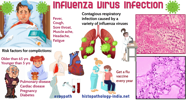
Orthomyxoviruses (influenza A, B, and C viruses) are enveloped viruses that contain single-stranded RNA in a helical array of nucleoprotein. Inserted in the lipid envelop are two glycoproteins - the hemagglutinin (HA) and the neuraminidase (NA). Influenza viruses have the unusual capacity of changing the antigenic identities of their HA and NA polypeptides, thus creating numerous antigenic variants. The host of different types of virus give their names to the disease, as in, for example, human, swine, and avian influenza. Avian Influenza (Bird Flu) Influenza viruses are highly contagious and afflict people of all ages. They are transmitted by aerosols generated by coughing and sneezing. Influenza A virus, the most common cause of viral pneumonia in adults, infects animals and man and produces pandemics. Several types of pneumonia associated with the influenza virus infection have been reported: 1) influenza complicated by secondary bacterial pneumonia, 2) primary influenza virus pneumonia, 3) combined influenza virus and bacterial pneumonia. Secondary bacterial pneumonia often produces a syndrome that is clinically distinguishable from that of primary viral pneumonia. Influenza B virus is apparently restricted to man, causes epidemics and is associated with Reye’s syndrome in children and pneumonitis and croup in infants. Influenza C virus causes sporadic respiratory infections, but not epidemic influenza. Influenza has significant mortality and morbidity, and may have long-term sequelae. Patients with viral influenza during the third trimester of pregnancy, the aged, and persons with valvular heart disease or chronic bronchopulmonary disease all have increased susceptibility to bacterial superinfection. Superinfection usually occurs 1 to 5 days after the onset of the vial illness, while the patient appears to be getting well. After an incubation period of 18 to 72 hrs, the onset is characteristically abrupt, with fever, chills, generalized malaise, myalgias and headache. As the fever and systemic symptoms subside, the respiratory symptoms become prominent. The histopathologic features include a necrotizing bronchitis and diffuse hemorrhagic necrotizing pneumonitis with pulmonary edema. Ciliated epithelial cells are destroyed and goblet cells and mucous glands disrupted. Bronchioles become thickened, distended, and infiltrated with mononuclear cells. There is often severe inflammatory edema, and the fluid exudates in the alveolar spaces has a hyaline appearance. In influenza significant life-threatening pathological conditions that could be considered the cause of death included diffuse alveolar damage, extensive secondary pneumonia, extensive intraalveolar hemorrhage, viral pneumonitis, myocarditis and meningoencephalitis.
|
|
|
