Custom Search


Dermpath-India

Pathology of Large Cell Acanthoma

Dr Sampurna Roy MD 


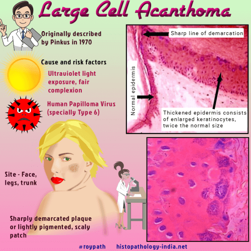
Large cell acanthoma usually presents as a well demarcated solitary plaque or scaly, lightly pigmented patch on sun exposed skin. 

HPV type 6 is considered an important cofactor in the pathogenesis of  large-cell acanthomas.

Site:  Usually located on the face, or on the limbs and trunk.


Microscopic features:

Histopathological examination reveals sharp demarcation of the lesion from the normal epidermis.

The thickened epidermis consists of enlarged keratinocytes, twice the  normal size.

Other features include basket weave orthokeratosis, prominent hypergranulosis and hyperpigmentation of basal layer.

In the dermis telangiectasia, solar elastosis  and chronic inflammation may be present.


Differential diagnosis includes solar lentigo and solar keratosis. 

(In solar keratosis there is parakeratosis, in large cell acanthoma there is orthokeratosis)


Further reading:

Large cell acanthoma: a debate throughout the decades

Large-cell acanthoma is a distinctive condition.

Clinicopathologic and immunohistochemical studies of conjunctival large cell acanthoma, epidermoid dysplasia, and squamous papilloma

Multiple disseminated large-cell acanthomas of the skin associated with human papillomavirus type 6.

Large cell acanthoma.

Large-cell acanthoma of the skin. A study by image analysis cytometry and immunohistochemistry.

Large-cell acanthoma is a solar lentigo.

Large-cell acanthoma of the eyelid. Report of two cases.


Visit:- Infectious Disease Online


Dr Sampurna Roy  MD

Consultant  Histopathologist (Kolkata - India)


Copyright © 2022  histopathology-india.net