

Custom Search
|

|
Infectious Disease Online Pathology of Leptospirosis
|
|
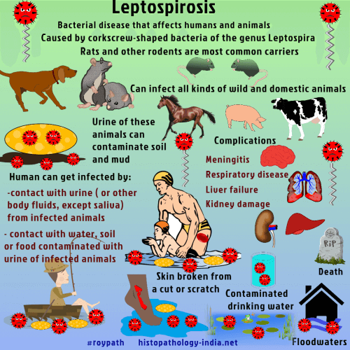
Leptospirosis is caused by the spirochete Leptospira interrogans. The leptospires are 0.1 micrometer wide and 6 to 12 micrometer long , with 18 or more coils. Of over 170 varieties, serovar canicola is associated with dogs, serovar icterohemorrhagica with rodents and serovar pomona with swine and cattle. Serovar icterohemorrhagica grows in the lumen of renal tubules in the rat and is shed in the urine for the life of the animal. The leptospires penetrate abraded skin or mucous membranes following contact with infected rats, contaminated water, or mud. Since worm, moist environments favour survival of the spirochetes, the incidence is greater in the tropics. Congenital infection causes fetal death. Symptoms begin 4 to 19 days after inoculation. Ninety percent of infections have a mild, anicteric course with resolution of symptoms in about 1 week. However, those with severe infections have a sudden onset of fever, myalgia , headache , and nausea and vomiting in the leptospiremic stage. The symptoms abate after 4 to 9 days and leptospires cease to circulate. The second, or immune stage known as Weil’s disease follows after a latent period of 1 to 3 days in 10% of patients. Fever, headache (which signals the onsetof meningismus), and the appearance of circulating IgM antibodies are characteristic. Severe myalgia, nausea, vomiting, abdominal pains, conjunctivitis, and hemorrhage into the conjunctiva are also features. Eventually hepatic failure, renal failure, and shock may lead to death. At autopsy there is a bile staining of tissues, hemorrhages in many organs, and pulmonary edema. Microscopically, the liver shows dissociation of the liver cell plates, erytrophagocytosis of the Kupffer cells, necrosis of hepatocytes, neutrophils in sinusoids, and a mixed inflammatory cell infiltrate in portal tracts. In the kidney the tubular epithelium is swollen and necrotic. Spirochetes are numerous in the lumen of the tubules and particularly in bile-stained casts. In the first phase, culture of blood and cerebrospinal fluid is the most effective means of confirming the diagnosis. PCR is a rapid, sensitive and specific means of diagnosing leptospiral infection, especially during the first few days of the disease. Leptospires grow from urine after the second week. Serologic tests are useful during the second phase. Antibiotics must be started within 4 days of onset. Large doses of penicillin and tetracycline are useful.
|
|
|
