

Custom Search
|


|
Dermpath-India Pathology of Intravascular Papillary Endothelial Hyperplasia (Masson's Tumour) Dr Sampurna Roy MD 2022
|
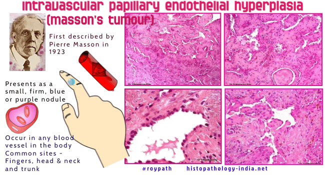
Intravascular papillary endothelial hyperplasia (Masson's tumour) was first described by Pierre Masson, who named it 'hemangioendotheliome vegetant intravasculaire' [Bull Soc Anat- (Paris) 1923; 93:517- 532]. Nine years later, Henschen described this proliferation as a reactive process rather than a true neoplasm and named the lesion ‘endovasculite proliférante thrombopoïétique’. In 1976, Clearkin and
Enzinger coined the current name ‘intravascular papillary endothelial
hyperplasia’, which has gained wide acceptance as the best description of
this lesion.
Site:
Masson's tumour may occur in any blood vessel in the body, but is commonly
located on the fingers, head and neck and trunk.
Less frequently, this
tumour has been documented in the upper respiratory tract,
skeletal muscle, urogenital systems, renal and hepatic systems, and
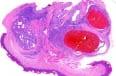
rarely in the gastrointestinal tract. Sectioning reveals cystic mass
containing clotted blood and surrounded by fibrous pseudocapsule. Microscopic Features : This is a well circumscribed lesion which is usually present within a blood vessel (commonly thin-walled vein). Multiple small, delicate papillary structures project into the lumen and these are associated with some thrombus. These papillae are lined by single layer of plump endothelial cells surrounding a collagenized core. There is no multilayering, tufting, solid areas, necrosis and little or no atypia. There is little evidence of mitoses. In the early lesions the the papillae are composed of fibrin. In the late stage there
is clumping and fusion of papillae forming an anastomosing network of
blood vessel set in a loose meshlike connective tissue. Unlike angiosarcoma, Masson's tumour is usually confined to the blood vessel (passive extension may occur following rupture of vessel) and there is no evidence of pleomorphism, tissue necrosis and mitoses. Masson's tumour is
typically positive for CD31, CD34, SMA, and factor VIII–related antigen.
CD105 is expressed on vascular endothelial cells and is positive in
primary vascular neoplasms, differentiating Masson's tumour from
angiosarcoma. Intravascular Papillary Endothelial Hyperplasia (Masson's Tumour) - Pathology Infographic
|
|
|

