|
|
|
||||
|
Dermatopathology Case No 28 (Images) 
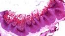
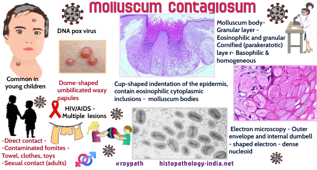
Molluscum contagiosum is benign viral disorder caused by a large brick-shaped DNA virus of the poxvirus group - Molluscum Contagiosum Virus. Age: May occur in all age groups. The disease is most commonly noted in young children (less than 5 years of age). May also occur in young adults due to sexual contact. Site: This is a disease of the skin and occasionally of the mucous membrane. Common sites include - Body, lower abdomen , arm, legs, inner thigh, buttock and genital area . Rare sites include eyelids ,lips and mouth.
Clinical presentation: Image Link Molluscum contagiosum, consists of a variable number of small discrete, waxy, skin-coloured, dome shaped papules. These are 2-4 mm in size with an umbilicated center. In a fully developed lesion a small amount of a curd-like substance can be expressed from the center. Occasionally a papule of molluscum contagiosum appears markedly inflamed. The lesion involute spontaneouly. In immunocompromised patients ( Eg:- AIDS ) hundreds of lesions of molluscum contagiosum are noted with little tendency to involute.
Microscopic features : ImageLink1 ; ImageLink2 ; ImageLink3 Epidermis grows down into the dermis as multiple, often closely packed lobules. Epidermal cells contain large, intracytoplasmic inclusion bodies, the so called molluscum bodies . Molluscum bodies first appear as single, minute ovoid eosinophilic structures at a level of two or three layers above the basal cell layer. Molluscum bodies increase in size as the infected cells move toward the surface. At the level of mid-epidermis the size exceeds the original size of the invaded cell . Molluscum bodies in the upper layers of the epidermis displace and compress the nucleus of each invaded cells. Nucleus appears as a thin crescent at the periphery of the cell. At the level of granular layer the staining reaction of molluscum bodies changes from eosinophilic to basophilic. In the centre of the lesion, stratum corneum ultimately disintegrates releasing the molluscum bodies together with keratinous debris , hence a central crater forms.
The surrounding dermis usually shows little or no inflammatory reaction except in rare instances in which the lesion of molluscum contagiosum ruptures and discharges molluscum bodies and keratinous debris. This may result in a prominent inflammatory infiltrate containing lymphoid cells, neutrophils, macrophages and in some cases a few foreign body giant cells. In lesions with follicular molluscum contagiosus , multiple greatly dilated hair follicles are present in the dermis filled with molluscum bodies.
|
January 2010
|
||||
Disclaimer ; Privacy Policy ; Advertising Policy ; E-mail
Copyright © 2010
histopathology-india.net
All rights reserved