Skin Appendage Tmour


http://www.histopathology-india.net/SwCa.htm

                          HISTOPATHOLOGY INDIA.COM                  

                  Path Quiz Case- 7 2

        Microcystic Adnexal Carcinoma

        Dr  Sampurna Roy  MD

                  Case history and images:

 
     DermPath-India

     Site created by

 Dr Sampurna Roy MD


http://www.histopathology-india.net/dermpath.htm

March 200
  Skin Appendage Tmour


http://www.histopathology-india.net/SAClass.htm

Benign sweat gland tumours

Cysts:

Hydrocystoma -Eccrine/Apocrine

Hamartomas:

Eccrine/Apocrine naevus

Eccrine Angiomatous Hamartoma

Porokeratotic eccrine ostial naevus

Benign Neoplasms:

Syringocystadenoma Papilliferum

Hidradenoma Papilliferum

Nipple Adenoma

Syringoma

Spiradenoma

Cylindroma

Chondroid Syringoma (benign mixed tumour)

Poroma

Hidradenoma

Apocrine adenoma

Papillary eccrine adenoma

Syringofibroadenoma

Malignant sweat gland tumour 

Low grade:

Microcystic adnexal carcinoma

Eccrine epithelioma

Adenoid cystic carcinoma

Mucinous carcinoma

Primary extramammary Paget’s disease

Intermediate grade:

Porocarcinoma

Digital papillary adenocarcinoma

High grade:

Hidradenocarcinoma

Apocrine adenocarcinoma

Eccrine ductal carcinoma

Spiradenocarcinoma

Malignant cylindroma

Sebaceous tumours

Fordyce's Spots

Steatocystoma

Nevus Sebaceous

Folliculosebaceous Cystic Hamartoma

Sebaceous Hyperplasia

Sebaceoma

Sebaceous Adenoma

Sebaceous Carcinoma

Tumours of the Hair Follicle

Hair Germ  Differentiation:

Trichoepithelioma 

Desmoplastic  Trichoepithelioma

Trichofolliculoma

Trichoblastoma

Cutaneous lymphadenoma


Infundibular differentiation:

Trichoadenoma

Dilated Pore of Winer

Pilar Sheath Acanthoma

Tumour of Follicular Infundibulum

Outer root sheath differentiation:
                    
Trichilemmoma

Trichilemmal Carcinoma

Proliferating Trichilemmal Cyst (Pilar Tumour)

Matrical differentiation:
                  
Pilomatrixoma and Pilomatrix Carcinoma

Histology of the Normal Skin

Gross examination of  the skin specimen

Reporting of biopsies taken for Inflammatory Skin Diseases

Lichenoid (Interface)Tissue  Reaction Pattern

Psoriasiform Reaction Pattern

Vesiculobullous Reaction Pattern

Spongiform Reaction Pattern

Vasculopathic Reaction Pattern

Lichen planus-like lesions

Lichen Nitidus

Bullous Pemphigoid

Hailey-Hailey Disease

Granuloma Annulare

Necrobiosis Lipoidica

Necrobiotic Xanthogranuloma

Rheumatoid Nodule

Foreign body granuloma


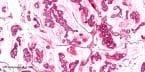
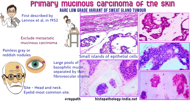
Primary mucinous carcinoma of the skin is a rare low grade variant of sweat gland tumour. 

Site:  Commonly arises in the head or neck, with the eyelid being the most common site. It is also found on the scalp, axilla and trunk.

Clinical presentation : Painless, gray or reddish nodules, measuring  0.5 to 7cm in diameter. Cut surface has a gelatinous appearance.

Microscopic features:  

Dermal tumour demonstrates large pools of basophilic mucin separated by thin fibrovascular septa ; Small islands of epithelial cells are present in the mucinous pool ; Cells are dense at the periphery; Tumour cells are small, cuboidal and bland ; May have vacuolated cytoplasm; Arranged in cribriform, papillary, tubular or glandular patterns ; Almost no mitotic figures are present.

Mucinous carcinomas originate from in situ lesions that may represent, ductal hyperplasia, atypical ductal hyperplasia, or ductal carcinoma in situ or a combination of the three. The presence of an in situ component defines the neoplasm as primary cutaneous, but its absence does not exclude the diagnosis.

Cytologic diagnosis of primary mucinous carcinoma of the skin is possible; however, correlation of clinical, radiologic and pathologic features is necessary to arrive at an accurate diagnosis.

Special stain: The mucin is PAS-positive and diastase resistant (consistent with sialomucin). Mucicarmine is also positive.

Immunohistochemistry:  Tumour is positive for low- molecular weight cytokeratin, CEA, EMA and sometimes S100.  Some cases demonstrated estrogen and progesterone receptors. CK20 is negative  which might significantly contribute to the differentiation of this tumour from cutaneous metastases from gastrointestinal carcinomas

In all cases of mucinous carcinoma of skin a full clinical assessment is essential. This tumour must be distinguished from metastatic visceral mucinous carcinoma (breast, colon, stomach, lung, ovary or pancreas). There should be no evidence of primary tumour at these sites.

Differential diagnosis:  Metastatic mucinous carcinoma ; Mucinous basal cell carcinoma - PAS negative

Colonic carcinoma metastatic to skin usually involves the anterior abdominal unlike primary cutaneous mucinous carcinoma which usually involves the head and neck area. Mucinous carcinomas of gastrointestinal origin produce nonsulfated, neutral and sulfated mucins . Primary mucinous carcinomas of the skin produce a nonsulfated mucin. Dirty necrosis is a constant histologic finding in most cases of intestinal mucinous carcinomas involving the skin

Mammary mucinous carcinoma involving the skin usually present with lesions on chest wall, breast, axilla and these locations can serve as clue to the breast origin.

Endocrine mucin-producing sweat gland tumour is an extremely rare tumour usually located on the eyelids. Endocrine mucin-producing sweat gland carcinoma: twelve new cases suggest that it is a precursor of some invasive mucinous carcinomas.Am J Surg Pathol. 2005;29(10):1330-9.
 

Treatment for primary mucinous carcinoma of the skin is wide local excision. Recurrence is common.  A few cases show metastatic spread to local lymph nodes. Mucinous carcinoma of the axilla is often associated with regional lymph node involvement.

            Image Link:(Dr Weems)    


 Abstracts of Articles: click here

Primary mucinous carcinoma of the skin. A case report.Ann Pathol. 2006 Jun;26(3):211-4

Fine needle aspiration biopsy of primary mucinous carcinoma of the skin: a case report.Acta Cytol 2006 May-Jun;50(3):317-22

Endocrine mucin-producing sweat gland carcinoma: report of a case and review of the literature.J Cutan Pathol. 2006 Dec;33(12):812-6.

Oestrogen and progesterone receptors in primary mucinous carcinoma of skin. Australas J Dermatol. 2005 ;46(4):246-9.

Primary cutaneous mucinous carcinoma of the eyelid in a young male.Orbit. 2005;24(3):211-4.

Primary Mucinous Carcinoma of the Skin: A Review. The Internet Journal of Oncology. 2005. Volume 2 Number 2.

Primary cutaneous mucinous carcinoma: presence of myoepithelial cells as a clue to the cutaneous origin.Am J Dermatopathol. 2004;26(5):353-8.

Mucinous carcinoma of the skin, primary, and secondary: a clinicopathologic study of 63 cases with emphasis on the morphologic spectrum of primary cutaneous forms: homologies with mucinous lesions in the breast.Am J Surg Pathol. 2005;29(6):764-82.

A rare case of fatal primary cutaneous mucinous carcinoma of the scalp with multiple in-transit and pulmonary metastases. J Am Acad Dermatol. 2005;52(5 Suppl 1):S76-80.

Primary cutaneous mucinous carcinoma initially diagnosed as metastatic adenocarcinoma. Tohoku J Exp Med. 2004;203 (4):345-8.

Mohs micrographic surgery of primary cutaneous mucinous carcinoma using immunohistochemistry for margin control. Dermatol Surg. 2004;30(5):799-802.

Secondary mucinous carcinoma of the skin: metastatic breast cancer. Dermatol Surg. 2004;30(2 Pt 1):234-5.

Primary mucinous carcinoma in a 54-year-old man.J Am Acad Dermatol. 2003 Nov;49(5):941-3.

Mucinous carcinoma of the skin with apocrine-type differentiation : immunohistochemical studies.Am J Dermatopathol. 2003 ; 25(1):66-70.

A case of primary mucinous carcinoma of the scalp treated with Moh's surgery.Dermatol Surg. 2002;28(8):751-4;discussion 754.

Primary mucinous carcinoma of the skin. American Journal of Dermatopathology. 2000; 22:166-170.

Primary mucinous carcinoma of the scalp. Dermatology. 2000;  200(3):250-3.

Long-standing translucent tumor on the groin. Diagnosis: mucinous carcinoma of the skin. Dermatology Online Journal. 1999; 5:4.2

Bone marrow relapse in primary mucinous carcinoma of skin.Am J Clin Oncol. 1999;22(3):303-4.

Primary eyelid mucinous adenocarcinoma of eccrine origin.Ophthalmic Surg Lasers. 1999;30(5):394-5.

Clinicopathological report: mucinous carcinoma of the eyelid.Aust N Z J Ophthalmol. 1999;27(1):71-3.

Primary mucinous carcinoma of the skin expresses TFF1, TFF3, estrogen receptor, and progesterone receptors. American Journal of Surgical Pathology. 1998; 22:1125-1131.

Primary mucinous carcinoma of the skin. Plastic and Reconstructive Surgery. 1997; 99:1160-1164.

Primary mucinous carcinoma of the skin. Report of a case. Pathologica. 1995; 87:689-691.

Mucinous carcinoma of the eyelid. An immunohistochemical study. American Journal of Dermatopathology. 1995; 17:494-498.

Sweat gland carcinoma with mucinous and infiltrating duct-like patterns.J Cutan Pathol. 1992;19(4):334-9.

Cytokeratin expression in mucinous sweat gland carcinomas: an immunohistochemical analysis of four cases. Histopathology. 1992 ;21(2) :161-5.

Primary mucinous carcinoma of the skin with metastases to the lymph nodes.Am J Dermatopathol. 1985 Oct;7(5):461-9.

Mucinous sweat gland adenocarcinoma of eyelid: a clinicopathologic study of 21 cases with histochemical and electron microscopic observations.Cancer. 1979;44(5):1757-68.

Your Banner


Histopathology-India.net

diagnostichistopathology. blogspot.com

Pathopedia-India.com

Surgical-Pathology.com

Pathology-India.com

Pancreatic Pathology Online

Gall Bladder Pathology Online

Paediatric Pathology Online

Paraganglioma-Online

Endocrine Pathology Online

Eye Pathology Online

Ear Pathology Online

Cardiac Path Online

Lung Tumour-Online

Mesothelioma-Online

Pulmonary Pathology Online

Nutritional Pathology Online

Environmental Pathology Online

Pathology Quiz Online

Dermpath-India

GI Path Online

Soft Tissue Pathology

Case Index

Infectious Disease Online; INDEX: A-D ; INDEX: E-L ; INDEX: M-P INDEX: Q-Z ; FUNGAL DISEASE ; VIRAL DISEASE.

E-book - History of  Medicine with special reference to India.

Basic Pathology Blog

Myxoid Tumours of Soft Tissue

Classification of Soft Tissue Tumour

Gross examination of soft tissue specimen          

A practical approach to histopathological reporting of soft tissue tumours

Grading of soft tissue tumours

Lipomatous tumours

Neural tumours

Myogenic tumours

Fibroblastic /Myofibroblastic tumours

Myofibroblastic tumours

Fibrohistiocytic tumours

ChondroOsseous tumours

Soft TissueTumours of Uncertain Differentiation               

Notochordal Tumour - Chordoma

Extra-adrenal Paraganglioma

Gastrointestinal Stromal Tumour

Cutaneous Pseudolymphoma

Drug related cutaneous lesions

Cutaneous Deposits

Calcinosis Cutis

Cutaneous Ossification (Osteoma Cutis)

Cartilaginous lesions of skin

Lipoid Proteinosis

Pathology of Colloid Milium

Gout

Silicone granulomas

Cutaneous Infections and Infestations

   1 : Bacterial, Rickettsial and Chlamydial Infections

   2 : Spirochetal Infections

   3 : Mycoses and algal Infections

   4 : Protozoal Infections

   5 : Helminth Infections

   6 : Viral Infections


                                                                           Copyright © 2009  histopathology-india.net
                                                                                              All rights reserved