Custom Search


Dermpath-India

Pathology of Nevoid Melanoma

Dr Sampurna Roy MD                                      2022
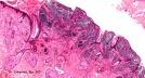
Dermatopathology Quiz Case: 183

Answer -   Nevoid melanoma


Nevoid melanoma is a rare form of nodular melanoma that may mimic benign melanocytic lesions. 

The lesions are called 'naevoid' because the cytological and architectural features resemble features of benign intradermal or compound melanocytic naevi.

Recent studies have shown that there is a significant local recurrence and mortality rate in patients diagnosed with naevoid melanoma.

Histopathological features:

To make a diagnosis of nevoid melanoma a high index of suspicion is essential.

An intraepidermal component is recognized at low power.

The intradermal component is composed of tightly packed nests of small dark stained cells resembling nevus cells.

The cells display hyperchromatic mildly pleomorphic nuclei, scanty cytoplasm and occasional prominent nucleoli.

Dermal mitosis may or may not be present. 

A reactive stromal response is evident characterized by proliferation of blood vessels and patchy lymphocytic infiltrate.

Some clues to diagnosis of Nevoid Melanoma: 

Hypercellularity ;

Loss of orderly arrangement of nests;

Cells present in strands and cords in some cases;

Presence of deep dermal mitosis ;

Lack of maturation with depth;

Infiltrative pattern at the base of the lesion.


Immunohistochemistry: 

S100 protein and HMB45  are usually positive.

The cells also express proliferating cell nuclear antigen (PCNA) and Ki67 (using MIB-1).

HMB45 may be negative in some cases.

Note:  In Spitz naevus there is stratification of staining .

In the base of the lesion cellular staining is less intense.

Similarities and differences between Nevoid melanoma and Compound naevus:

Nevoid malignant melanoma small cell type:

Similarities:

i) Cells are usually small ;

ii) Pagetoid cells are rare ;

iii) Melanocytes are nested

iv)Cells may disperse at the base.

Differences:

Compound Naevus:  Mitosis is absent;  Nucleoli inconspicuous at base of the lesion; Nuclei not hyperchromatic ; HMB45 negative at base; MIB1 negative at base.

Naevoid malignant melanoma (small cell type): Mitosis present in dermis ; Nucleoli visible at the base; Nuclei hyperchromatic ; HMB45- positive at base;  MIB-1 positive at base.


Similarities and differences between Nevoid melanoma and Spitz's Nevus:

Nevoid Malignant Melanoma Large Cell Type:

Similarities:

(i) Cells tend to be large;

(ii) Pagetoid cells may be present;

(iii) Melanocytes are nested

(iv) Cells may disperse at the base.

Differences:

Spitz's naevus :

Mitoses present in dermis, but rarely at the base; cell size small  at the base;  Kamino bodies are common; HMB-45 patchy positivity, may be at base ; MIB-1 diminishes at base.

Nevoid malignant melanoma (large cell type) :

Mitoses present in dermis even at base; Cell size large at base ; Few Kamino bodies; HMB-45 positive at base; MIB-1 undiminished at base.

Verrucous Nevoid Melanoma:

Melanoma is characterized by epidermal hyperplasia , elongation of rete ridges  and overlying hyperkeratosis.

Clinically these lesions may be mistaken for seborrheic keratosis.

(At low power some these lesions may resemble papillomatous benign  intradermal naevi.)

The tumour consists of small naevus like cells with no evidence of maturation  with depth.

The lesion may demonstrate atypical junctional component, asymmetric lateral growth pattern and area of regression.


Further reading:

Morphological analysis of nevoid melanoma. A study of 20 cases with a review of literature. 

Small cell malignant melanoma: a variant of naevoid melanoma. Clinicopathological features and histological differential diagnosis.

"Triggered trap"- nevoid malignant melanoma. 

Small cell melanoma: A clinicopathologic study of 131 cases. 

Small cell (naevoid) melanoma: a clinicopathologic study of 131 cases. 

Nevoid melanoma: a clinicopathological study of seven cases of malignant melanoma. mimicking spindle and epithelioid cell nevus and verrucous dermal nevus. 

Nevoid malignant melanoma: morphologic patterns and immunohistochemical reactivity

Verrucous naevoid and keratotic malignant melanoma: a clinico-pathological study of 20 cases.
                                                              
Verrucous-keratotic variations of malignant melanoma. A clinicopathological  study. 

Verrucous pseudonaevoid melanoma


Visit:- Infectious Disease Online

Dr Sampurna Roy  MD

Consultant  Histopathologist (Kolkata - India)


Copyright © 2022  histopathology-india.net