

Custom Search
|


|
Dermpath-India Pathology of Ossifying Fibromyxoid Tumour Dr Sampurna Roy MD 2022
|
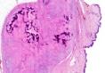

| Ossifying
fibromyxoid tumour (OFMT) is a rare soft tissue tumour of uncertain
histogenesis. The tumour presents as a well circumscribed small subcutaneous or muscular nodule on the extremities. Ossifying fibromyxoid tumor in soft parts was described by Enzinger et al. in 1989. Although the tumour most commonly occurs on the extremities with a male predominance, cases of ossifying fibromyxoid tumour have been reported in females, on the scalp (occurring as a cystic lesion) , the nasal septum (history of nasal airway obstruction and enlargement contour of the nose) and in the orbit (history of diplopia, pain, and right upper eyelid swelling). This is an indolent tumour, although local recurrences may occur.
Line of
differentiation of this tumour is uncertain, however it has been suggested
that the tumour is probably of partial neural, myoid or even of
myofibroblastic origin. Microscopically, on the low power, the tumour is lobulated and an incomplete shell of mature bone is note in the area corresponding to the fibrous septa. The tumour consists of lobules of uniform rounded cells arranged in cords or strands. These cells are set in a fibromyxoid stroma. Mitotic figures are inconspicuous. Unusual features include satellite micronodule formation, multiple microcalcifications, epidermoid cysts and mucinous microcyst formation, absence of myxoid areas, foci of atypical chondroid differentiation, binucleate lacunar cells, pericytic growth pattern and malignant change. Atypical or malignant ossifying fibromyxoid tumour has been described by Kilpatrick et al. The lesion is characterized by increased cellularity and prominent mitotic activity. Some of these cases may metastasize to the lungs. Despite showing benign characteristics, these tumors can present aggressive behavior, and they should not therefore be classified as benign, but rather considered as tumors of intermediate malignancy. Immunohistochemistry reveals that the tumour is positive with S100 protein and vimentin. Many cases are desmin positive. Smooth muscle actin is positive in about 50% cases. There is also focal reactivity for Leu7 and GFAP. Cytogenetic analysis of this type of tumor focuses attention on 2 genes, INI-1, a tumor suppressor gene, and PHF1, which codes for a protein that, among other functions, regulates the activity of the polycomb-repressive complex 2, which silences genes responsible for development.
|
|
|

![]()
Copyright © 2002-2022 histopathology-india.net