Gastrointestinal Stromal Tumour


http://www.histopathology-india.net/SoftTissuePath.htm

                                HISTOPATHOLOGY INDIA.COM

                       Neurofibroma

           Dr Sampurna Roy MD
 
Web www.histopathology-india.net

 June 2007
Normal Anatomy of the Nerve

Surgical-Pathology.com

Histopathology-India.net

Eye Pathology Online

Cardiac Path Online;

Pulmonary Pathology Online

Pathology Quiz Online;

Dermpath-India;

GI Path Online

Mesothelioma-Online;

Soft Tissue Pathology;

Case Index

Infectious Disease Online; INDEX: A-D ; INDEX: E-L ; INDEX: M-P INDEX: Q-Z ; FUNGAL DISEASE ; VIRAL DISEASE.

Reactive and hamartomatous lesions:

Traumatic neuroma

Morton's neuroma

Digital Pacinian neuroma

Nerve Sheath Ganglion

Fibrolipomatous hamartoma of nerve

Benign tumours:

Solitary circumscribed neuroma
(palisaded encapsulated neuroma)

Schwannoma (neurilemmoma)
andvariants                          

Neurofibroma and variants 

Perineurioma   

Dermal nerve sheath myxoma
(neurothekeoma)

Cellular neurothekeoma

Granular cell tumour

              
Malignant tumour:

Malignant peripheral nerve sheath tumour


Neuroendocrine Carcinoma:

Merkel cell (neuroendocrine) carcinoma

Malignant primitive neuroectodermal tumour   

Miscellaneous neuroectodermal tumours presenting in soft tissue

Subcutaneous Myxopapillary Ependymoma  

Heterotopic Glial Nodule

Heterotopic Meningeal Lesions

Malignant peripheral nerve sheath tumours may show perineurial cell differentiation.These tumours are epithelial membrane antigen positive and  S-100 protein negative. The prognosis of perineural MPNST appears to be more favorable than that of conventional MPNST.
Myxoid Tumours of Soft Tissue

Classification of Soft Tissue Tumour

Gross examination of soft tissue specimen          

A practical approach to histopathological reporting of soft tissue tumours

Grading of soft tissue tumours

Lipomatous tumours

Neural tumours

Myogenic tumours

Fibroblastic/Myofibroblastic tumours

Myofibroblastic tumours

Fibrohistiocytic tumours

ChondroOsseous tumours

Soft TissueTumours of Uncertain Differentiation               

Notochordal Tumour - Chordoma

Extra-adrenal Paraganglioma

Gastrointestinal Stromal Tumour


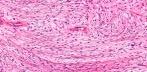
Peripheral nerves are surrounded by an external sheath, which contains concentric layers of thin perineurial cells. Perineuriomas (PN) are rare nerve sheath tumours composed of well-differentiated perineurial cells exhibiting immunoreactivity for epithelial membrane antigen (EMA). These lesions occur preferentially in adults and may arise in a wide variety of anatomic sites.

3 main forms of  perineurioma:

1. Extraneural Soft tissue PN including a conventional form: This is the common type and presents as a painless subcutaneous  mass on the limbs or trunk of middle aged adults.

2. Intraneural PN: Rare type. Presents as a swelling of a major nerve in young adults.

3. Sclerosing perineurioma: Solitary, small nodule in the finger or palm usually in young adults and is associated with deletion of NF2 gene on chromosome 22.

  Other variants:  Fibrous and reticular perineuriomas

Microscopic features:

(ESCOP)IMAGE LINKS1   2   3   4       CaseLink (Spain)

Well circumscribed non-encapsulated lesion composed of spindle cells with eosinophilic cytoplasm and elongated nuclei. The cells are parallel to each other or form small concentric whorls (onion bulbs).

All perineuriomas are EMA positive (focally in some cases), Claudin-1 positive and S100 protein negative.

Common electron microscopic features are organelle-poor cell processes, many pinocytotic vesicles, sparse intermediate filaments, tight junctions and patchy external lamina.

Sclerosing perineurioma:   Well-circumscribed lesion characterized by small epithelioid cells exhibiting corded, trabecular and whorled growth patters together with spindle cells with wavy nuclei end elongated cytoplasmic processes and thick collagen. There is focal whorling of tumour cells. The cells are set in a dense collagenous backround. Immunohistochemically, most of the tumour cells are positive for epithelial membrane antigen, vimentin, collagen type IV and CD10, but not for S-100 protein, CD34, desmin and cytokeratin. The tumor cells are positive for the human erythrocyte glucose transporter (GLUT1) antigen suggesting that this could be an useful marker for the identification of sclerosing perineurioma.

Reticular perineurioma:   Microscopically the lesions are characterized by a predominantly lace-like or reticular growth pattern composed of anastomosing cords of fusiform cells with bipolar cytoplasmic processes and palely eosinophilic cytoplasm. Nuclei were centrally placed, ovoid to fusiform in shape, and no mitoses were seen. Transition to more cellular areas was focally present in all cases. D/D: Myoepithelial tumors, extraskeletal myxoid chondrosarcoma, and myxoid synovial sarcoma.

Cutaneous fibrous perineurioma:
  These tumours presented with sharp circumscription of their deep aspect or were not circumscribed on any side. The cells included plump and spindled with one or more nuclei to  thin, elongated, spindled cells with slender nuclei. The backround stroma is fibrotic. Immunohistochemical staining showed EMA-positive staining of the cellular component  with collagen type IV-positive staining surrounding the cells.The tumour cells were negative for S-100 protein, factor XIIIa, CD34 and cytokeratin.

Perineuriomas may arise in the intestine usually as intramucosal lesions detected as colorectal polyps with distinctive histologic features including entrapment of colonic crypts. Intestinal  perineuriomas are distinguished from other spindle cell neoplasms of the gastrointestinal tract by immunostaining for EMA and claudin-1.

Soft tissue perineuriomas :  Behave in a benign fashion and rarely recur. Atypical histologic features (including scattered pleomorphic cells and infiltrative margins) seem to have no clinical significance.


Web www.histopathology-india.net
Abstracts:

Soft tissue perineurioma: clinicopathologic analysis of 81 cases including those with atypical histologic features. Am J Surg Pathol. 2005 Jul;29(7):845-58.

Intestinal perineuriomas: clinicopathologic definition of a new anatomic subset in a series of 10 cases.Am J Surg Pathol. 2005 Jul;29(7):859-65

Perineurioma: a clinicopathological study of eight cases.Pathology. 2004 Aug;36(4):309-15

Soft tissue perineurioma: evidence for an abnormality of chromosome 22, criteria for diagnosis, and review of the literature. Am J Surg Pathol 1997;21:164–73

Cutaneous sclerosing perineurioma of the digits: an uncommon soft-tissue neoplasm. Report of two cases with immunohistochemical analysis. J Cutan Pathol. 2003 Oct;30(9):577-581

Sclerosing perineurioma: a clinicopathological study of five cases and diagnostic utility of immunohistochemical staining for GLUT1.Virchows Arch. 2003 Aug;443(2):159-63. Epub 2003 Jun 26.

Expression of claudin-1, a recently described tight junction-associated protein, distinguishes soft tissue perineurioma from potential mimics.Am J Surg Pathol 2002 ;26 (12): 1620-6

Sclerosing perineurioma: a clinicopathologic study of 19 cases of a distinctive soft tissue lesion with a predilection for the fingers and palms of young adults. Am J Surg Pathol. 1997 Dec;21(12):1433-42.

Perineurioma of the kidney. Report of a case with histologic, immunohistochemical, and ultrastructural studies. Arch Pathol Lab Med 1993 Jun;117(6):654-7

The Clinical and Histologic Spectrum of Cutaneous Fibrous Perineuriomas .Am J Dermatopathol 2001;23:190-196

Cutaneous fibrous perineurioma. J Cutan Pathol 1998;25:333–7.

Perineuroma. A frequently unrecognized entity with emphasis on a plexiform variant.Adv Clin Path. 2000 Jan;4(1):25-33

Reticular Perineurioma A Distinctive Variant of Soft Tissue Perineurioma. Am J Surg Pathol 2001;25:485-493

Cutaneous perineurioma. Clinical and histological findings and differential diagnosis.Pathologe. 2003 May;24(3):207-13. Epub 2003 Mar 19

Malignant peripheral nerve sheath tumor with perineurial differentiation: 'malignant perineurioma'. J Cutan Pathol 2002 Jul;29(6):362-7

Perineurial malignant peripheral nerve sheath tumor (MPNST): a clinicopathologic, immunohistochemical, and ultrastructural study of seven cases.Am J Surg Pathol. 1998 Nov;22(11):1368-78.

Soft tissue perineurioma of the finger: expanding the differential diagnosis of a soft tissue tumor presenting on a digit.Cutis. 2005 Apr;75(4):233-7.

Soft tissue perineurioma in a patient with neurofibromatosis type 2: a tumor not previously associated with the NF2 syndrome.Am J Surg Pathol. 2006 Dec;30(12):1624-9.

Benign hybrid perineurioma-schwannoma in the colon. A case report.
Ann Diagn Pathol. 2006 Dec;10(6):367-70.

Hybrid peripheral nerve sheath tumors: Schwannoma-perineurioma and neurofibroma-perineurioma. A report of three cases in extradigital locations.Ann Diagn Pathol. 2005;9(1):16-23.

Perineurioma: an uncommon soft tissue neoplasm distinct from localized hypertrophic neuropathy and neurofibroma.Am J Surg Pathol 1992;16(8):756-63

Low-grade malignant perineurioma of the paravertebral column, transforming into a high-grade malignancy. Pathol Int 1999 ;49(9):820-5

Perineurial cell tumor (perineurioma) with granular cells. J Cutan Pathol 1997;24(9):575-9

Immunohistochemical detection of epithelial membrane antigen in normal perineurial cells and perineurioma.Am J Surg Pathol. 1988 Sep;12(9):678-83.

 Cardiac Path Online;

PRIMARY TUMOURS OF THE HEART ; REPORTING OF CARDIAC TUMOURS ; CARDIAC MYXOMA ;CARDIAC RHABDOMYOMA ; PAPILLARY FIBROELASTOMA ; CARDIAC FIBROMA ; CARDIAC LIPOMA ;CARDIAC HEMANGIOMA ; CARDIAC TERATOMA ; MESOTHELIOMA OF ATRIOVENTRICULAR NODE ;PURKINJE CELL TUMOUR ; CARDIAC PARAGANGLIOMA ; MALIGNANT TUMOURS OF THE HEART ; CARDIAC LYMPHOMA ;