Custom Search


Dermpath-India

Pathology of Pilomatrixoma

Dr Sampurna Roy MD                                          2022


Dermatopathology Quiz Case: 252

Answer -  Proliferating Pilomatrixoma


Dermatopathology Quiz Case: 208

Answer - Pilomatrixoma


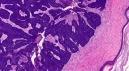
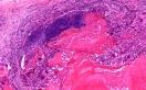
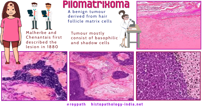
Syn:  Pilomatricoma ; Calcifying epithelioma of Malherbe

Pilomatrixoma is a very common benign adnexal tumour with matrical differentiation.

Both ultrastructurally and histochemically these are similar to hair matrix.

Most cases are associated with beta-catenin gene mutations.

Clinical presentation: 

Solitary and rarely multiple rubbery nodules (usually 5mm to 2cm in diameter).

The tumour usually occurs in the first two decades of life. Often diagnosed as epidermoid/pilar cysts. 

May be associated with Gardner's syndrome or myotonic dystrophy. Usually multiple lesions are present.

Site: Located on the head, neck or forehead .


Microscopic features:

- Well circumscribed lobular sometimes cystic tumours ;

- Composed of proliferating basaloid (resemble matrical cells of the  follicular bulb) and eosinophilic shadow keratinised cells (represent attempted cortical and inner root sheath differentiation) ;

- Dystrophic calcification and giant cell reaction to keratin and shadow cells;

- Sometimes osseous metaplasia may be present ; 

Cystic pilomatrixoma shows more basaloid cells. Solid lesions are usually devoid of basaloid cells and there are more shadow cells, calcification and giant cell reaction.


Proliferating Pilomatrixoma:

A distinctive proliferative variant of pilomatrixoma ; 

This is a relatively large lesion in the dermis which sometimes extend to the  subcutis ;

Microscopic features:

Composed of lobular proliferation of basaloid cells in association with adjacent focal areas containing eosinophilic, cornified material with shadow cells ; 

In some cases there are relatively large areas of shadow cells whereas, in others there are only small foci of shadows cells ;

Basaloid cells show variable nuclear atypia and mitotic figures ;

Architectural pattern of the neoplasms is different from that of a large fully developed pilomatrixoma with a cystic character and basaloid cells predominantly aligned at the periphery. 

Differential diagnosis: Matricoma, basal-cell carcinoma with matrical differentiation, and matrical carcinoma (pilomatrical carcinoma).

The anetodermal or lymphangiectatic variant of pilomatricoma is rare, and its bullous appearance is often associated with attenuated collagen and elastic fibrils and dilated lymphatic vessels in the overlying dermis.


Further reading:

Pilomatrixoma - Presented as Hypopigmented Tender Nodule: Diagnosed by FNAC: A Case Report with Review of Literature.

Cytodiagnosis of pilomatrixoma from an uncommon site with unusual presentation

Multiple pilomatricomas with somatic CTNNB1 mutations in children with constitutive mismatch repair deficiency.

Pilomatrixoma of the arm: a rare case with cytologic diagnosis.

Pilomatricoma: an unusual dermatologic neoplasm.

Pilomatrixoma: an infrequently encountered lesion on the lower extremity.

Epithelioma of Malherbe: new ultrasound patterns.

Pilomatricoma of the arm with extensive ossification.

Additional cytomorphological criteria in diagnosis of pilomatricoma--benign tumor with bad reputation.

Multiple pilomatricomas: case presentation and review of the literature.

Immunohistochemical evaluation of inflammatory reaction in a case of pilomatrixoma.


Dr Sampurna Roy  MD

Consultant  Histopathologist (Kolkata - India)


Copyright © 2002-2022  histopathology-india.net