DermPath-India

     Site created by

 Dr Sampurna Roy MD


http://www.histopathology-india.net/dermpath.htm


                          HISTOPATHOLOGY INDIA.COM

            Myxoid Tumours of Soft Tissue

   Dr Sampurna Roy MD

 
   Gastrointestinal Stromal Tumour


http://www.histopathology-india.net/SoftTissuePath.htm

January 2008 
Surgical-Pathology.com

Histopathology-India.net

Pathology-India.com

Pancreatic Pathology Online

Gall Bladder Pathology Online

Paediatric Pathology Online

Paraganglioma-Online

Endocrine Pathology Online

Eye Pathology Online

Ear Pathology Online

Cardiac Path Online

Lung Tumour-Online

Mesothelioma-Online

Pulmonary Pathology Online

Nutritional Pathology Online

Environmental Pathology Online

Pathology Quiz Online

Dermpath-India

GI Path Online

Soft Tissue Pathology

Case Index

Infectious Disease Online; INDEX: A-D ; INDEX: E-L ; INDEX: M-P INDEX: Q-Z ; FUNGAL DISEASE ; VIRAL DISEASE.

Cutaneous infection and infestations

Histopathological patterns in cutaneous infections

1: Bacterial, Rickettsial and Chlamydial infection

2 : Spirochetal Infection

3 : Mycoses and algal infections

4 : Protozoal Infections

5 : Helminth Infections

6 : Viral Infections

Cutaneous lesion associated with AIDS

Granulomatous Reaction Pattern of the Skin

Granuloma Annulare

Necrobiosis Lipoidica

Necrobiotic Xanthogranuloma   

Rheumatoid Nodule

Lupus Vulgaris

Cutaneous Sarcoidosis

Melkersson Rosenthal Syndrome

Annular Elastolytic Giant Cell Granuloma

Skin lesion in Crohn's Disease

Blastomycosis-like pyoderma


 Microscopic  Image

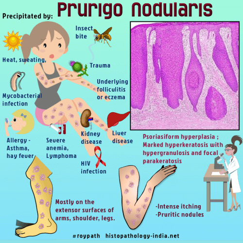
Prurigo nodularis is an uncommon lesion that presents as extremely pruritic nodules with a  wide anatomical distribution but particularly on extensor aspects of limbs.

Often lesions are multiple but solitary nodules can occasionally occur.

Lesions tend to be symmetrical and are associated with excoriations.DermAtlas Images

Females are more often affected than males.

Many authors consider this condition as part of the spectrum of lichen simplex.

Atypical mycobacteria may be a contributing factor in prurigo nodularis.

Microscopic features: 

Histopathology reveals psoriasiform hyperplasia or pseudo- epitheliomatous hyperplasia  ;

Marked hyperkeratosis with hypergranulosis and focal parakeratosis ;

Occasional spongiosis and exocytosis of mononuclear cells;

Scale crust formation in association with changes of excoriation (fibrin deposition and cellular debris) ;

Prominent hyperplasia of hair follicles;

Mild fibosis of papillary and superficial dermis and telangiectasia ;

Mild to moderate perivascular mononuclear inflammatory cell infiltrate composed of lymphocytes , histiocytes and plasma cells ;

Nerve hyperplasia as reported in the past is not usually a feature.

Differential diagnosis:  Diagnosis is not difficult if the biopsy is big enough. In small biopsies differential diagnosis include Keratoacanthoma  and other causes of pseudo- epitheliomatous hyperplasia.

Note:   Pseudoepitheliomatous (pseudocarcinomatous) hyperplasia :  This is a benign pathological reaction pattern, histologically  characterized by bulbous thickening of squamous epithelium (follicular infundibula and acrosyringium). There is abrupt transition  beween lesion and adjacent epidermis. The cells have abundant often glassy cytoplasm .
Pseudoepitheliomatous hyperplasia is seen in the  following conditions:
Chronic irritation ;  skin trauma ;  dermal inflammatory processes like chromomycosis ; aspergillosis ;  pyoderma,
Prurigo nodularis ; Granuloma fissuratum and in associatiation with chondrodermatitis nodularis helicis ;  Spitz naevus , melanoma,  overlying granular cell tumour and cutaneous T cell tumour .


Abstracts:

Prurigo nodularis: a review.Australas J Dermatol. 2005 Nov;46(4):211-18; quiz 219-20.

Increased nerve growth factor- and tyrosine kinase A-like immunoreactivities in prurigo nodularis skin -- an exploration of the cause of neurohyperplasia.Arch Dermatol Res. 2002 Feb;293(12):614-9. Epub 2002 Feb 5.

Eosinophil cationic protein- and eosinophil-derived neurotoxin/eosinophil protein X-immunoreactive eosinophils in prurigo nodularis.Arch Dermatol Res. 2000 Aug;292(8):371-8.

Light and electron microscopic immunohistochemical observations of p75 nerve growth factor receptor-immunoreactive dermal nerves in prurigo nodularis.Arch Dermatol Res. 1999 Jan;291(1):14-21.

Helicobacter pylori and prurigo nodularis..Hepatogastroenterology. 1999 Jul-Aug;46(28):2269-72.

Histamine-containing mast cells and their relationship to NGFr-immunoreactive nerves in prurigo nodularis: a reappraisal.J Cutan Pathol. 1998 Apr;25(4):189-98.

Syringomatous changes of eccrine sweat ducts associated with prurigo nodularis.Am J Dermatopathol. 1998 Jun;20(3):296-301

Histopathological and bacteriological findings in prurigo nodularis.Acta Derm Venereol. 1997 Jan;77(1):49-51.

Mycobacteria in prurigo nodularis: the cause or a consequence?J Am Acad Dermatol. 1996 Feb;34(2 Pt 1):224-8.

Merkel cells and prurigo nodularis. J Am Acad Dermatol. 1994 Jul;31(1):86-8.

Mast cells, neutrophils, and eosinophils in prurigo nodularis. Arch Dermatol.1993 Jul;129(7):861-5.

Demonstration by S-100 protein staining of increased numbers of nerves in the papillary dermis of patients with prurigo nodularis.J Am Acad Dermatol. 1992 Jan;26(1):56-8.


Epidermal tumours:

Epidermal Naevus ; Inflammatory linear verrucous epidermal nevus

Prurigo Nodularis

Acanthomas - [ Epidermolytic acanthoma;Acantholytic acanthoma; Melanoacanthoma ]

Clear cell acanthoma

Large cell acanthoma

Warty Dyskeratoma

Seborrheic Keratosis

Verruca vulgaris; Palmoplantar wart; Verruca plana ; Epidermodysplasia verruciformis ; Condyloma acuminatum

Keratoacanthoma

Actinic Keratosis

Bowen's disease

Basal Cell Carcinoma

Squamous Cell Carcinoma

Cutaneous Squamous Cell Carcinoma (Image &abstracts)

Normal Histology of Skin

Glossary   

Gross examination of the skin specimen

Reporting of biopsies taken for Inflammatory Skin Diseases

Lichenoid (Interface)Tissue Reaction Pattern

Psoriasiform Reaction Pattern

Vesiculobullous Reaction Pattern

Spongiform Reaction Pattern

Vasculopathic Reaction Pattern

Lichen planus-like lesions

Bullous Pemphigoid

Dermatitis Herpetiformis

Hailey-Hailey Disease

Erythema Nodosum

Folliculitis

Impetigo

Furuncle(Boil)

Carbuncle

Toxic Shock Syndrome

Drug related cutaneous lesions

Cutaneous lesion in graft-versus host disease

Verruciform Xanthoma

Xanthelasma

Cutaneous Deposits

Calcinosis Cutis

Cutaneous Ossification (Osteoma Cutis)

Cartilaginous lesions of skin

Lipoid Proteinosis

Pathology of Colloid Milium

Gout

Silicone granulomas

Foreign body granuloma

Rosacea

Interstitial Granulomatous Dermatitis

Interstitial Granulomatous Drug Reaction

Granulomatous T-cell lymphoma