Custom Search

Dermpath-India

Pathology of Pigmented Spindle Cell Nevus (Reed Nevus)

Dr Sampurna Roy MD                 2022

Dermatopathology Quiz Case: 185

Answer - Pigmented Spindle Cell Nevus (Reed Nevus)


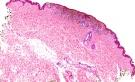
Reed nevus or pigmented spindle-cell nevus was first described by Reed in 1975.

Pigmented spindle cell nevus is regarded as a distinct entity and not part of spindle cell variant of Spitz nevus.

Clinically, these lesions present as symmetrical, sharply circumscribed, darkly pigmented nodule or papule (usually less than 0.6 cm in diameter).  

These lesions occur most commonly on the extremities and back with a predilection for the legs.

These are more common in women in the third decade of life.


Microscopic features:

- Histologically, the tumour consists of spindle shaped cells arranged in nests with formation of interconnecting fascicles.   

- Prominent vertical orientation of cells are noted (Raining down appearance ).

- The tumour has a expansile growth pattern.

- It is heavily pigmented and melanophages are present.

- Kamino bodies (similar to Spitz) are noted in pigmented spindle cell nevus.

- Junctional clefting is noted in some cases.


Differential diagnosis include melanoma.

Pigmented spindle cell nevus  that can show worrisome clinical and histologic features mimicking a malignant melanoma.

Unlike melanoma these are symmetrical lesions and display maturation of cells in the deeper part of the lesion.

Pagetoid spread of cells is limited to the lower half of the epidermis.  

Clinical and histologic assessment is essential in establishing the diagnosis

Ancillary techniques such as proliferation antigen Ki-67, cyclin D1, survivin, and FISH can be useful as adjunctive tools. 

Dermatopathology Quiz Case 135 

[Pathology Infographic] Raining down appearance of Pigmented Spindle Cell Nevus (Reed Nevus)


Further reading:

Pigmented spindle-cell nevus: A melanoma simulator.

Pigmented spindle cell naevus of reed presenting in the conjunctiva.

Pigmented spindle cell nevus: a clinicopathologic analysis of ninety-five cases.

The histologic spectrum of pigmented spindle cell nevus: a review of 120 cases with emphasis on atypical variants.

The pigmented spindle cell tumor of Reed: an underdiagnosed lesion.

Pigmented spindle cell nevus.

Pigmented spindle cell naevus of Reed presenting in the conjunctiva

Eosinophilic globules in pigmented spindle cell nevus

Pigmented spindle cell nevus: clues for differentiating it from spindle cell malignant melanoma.

Pigmented spindle cell nevus: clues for differentiating it from spindle cell malignant melanoma. A comprehensive survey including clinicopathologic, immunohistochemical, and FISH studies.


Visit:- Infectious Disease Online

Dr Sampurna Roy  MD

Consultant  Histopathologist (Kolkata - India)


Copyright © 2022  histopathology-india.net