

Custom Search
|


|
Pathology Sebaceous Adenoma Dr Sampurna Roy MD 2022
|

 |
|
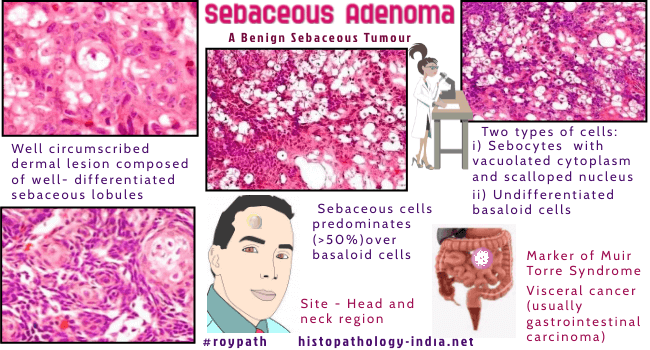
Sebaceous adenoma is a benign sebaceous tumour. It is closely associated with the Muir-Torre syndrome. Clinical presentation: May be solitary or multiple ; Yellow-pink circumscribed papule/nodule (0.5 cm in diameter) ; This may ulcerate or bleed. Site: Located on the head and neck region and rarely on the buccal mucosa. Microscopic features: Histologically, sebaceous adenoma is a multilobulated tumour sharply demarcated from the surrounding tissue. Two types of cells are present in the lobules: The large mature sebaceous cells (sebocytes) are present at the centre. Smaller, undifferentiated basaloid cells in the periphery. The cellular lobules contain ductal strutures with holocrine secretion. Sometimes lobules contain cystic spaces in the centre due to disintegration of mature sebaceous cells.
|
|
|
|
|
