|
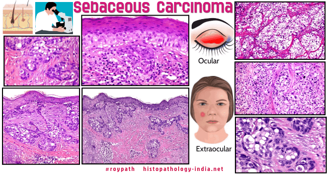
Sebaceous carcinoma is an aggressive tumour with both local recurrence
and distant metastasis.
It has been noted
that this lesion commonly occurs in the Asian population.
Sebaceous
carcinoma is rarely associated with Muir-Torre syndrome.
These tumours have been
divided into 2 groups : Ocular and Extraocular.
Ocular sebaceous
carcinoma is more common.
Ocular cases may mimic chalazion.
The tumour arises from the glands in the periocular region (Meibomian
glands, Glands of Zeis).
Extraocular
sebaceous carcinoma is usually noted in the head and neck area.
Grossly, the tumour
presents as nodules, papules and plaques.
| Microscopic features:
Histology reveals irregular lobular growth pattern composed of
sebaceous and undifferentiated cells.
These cells show variation in the shape and size of nuclei.
The undifferentiated cells show eosinophilic cytoplasm with fine lipid
globules.
Some large lobules show areas of atypical keratinizing cells.
There is pagetoid spread of malignant cells in the conjunctival
epithelium or epidermis of the skin of the lid.
Differential Diagnosis-
Extramammary Paget's disease:
This is rarely seen in extraocular sebaceous carcinoma.
The pagetoid cells contain no mucopolysaccharides but stain positively
for fat with oil red O .
[NOTE:
Identification of pagetoid
growth pattern in biopsy material is essential to recognize the
presence of an underlying sebaceous carcinoma.
]
Special stains:
Image
To highlight the presence of tumour cells with sebaceous
differentiation one would require special histochemical techniques,
such as oil red O,
Sudan IV stains on fresh tissue and EMA immunostains on paraffin
embedded tissue.
|
Differential diagnosis:
Squamous cell
carcinoma ; Basal cell carcinoma with sebaceous differentiation.
Pathology of
Sebaceous Carcinoma [Infographic]
|
Criteria for Malignancy in Sebaceous Neoplasia:
Lazar AJF, Lyle S, Calonje E. Sebaceous
neoplasia and Torre–Muir syndrome. Current diagnostic pathology
2007;13(4):301-319.
-
Sebaceous
tumors showing an infiltrative border or cytologic atypia should
regarded as malignant.
- Tumor
necrosis in basaloid areas is a strong indicator of malignancy,
but should not be confused with areas of holosecretion in
sebaceous adenomas.
- Mitoses are
not a strict criteria for malignancy as sebaceomas can sometimes
show conspicuous mitotic activity. An unusual number of mitoses
or atypical mitotic forms must be interpreted within
the overall appearance of the tumor.
-
Pagetoid
intraepithelial migration indicates malignancy, but is not
commonly encountered in non-ocular cases.
-
All sebaceous
tumors of the eyelid should be regarded as malignant unless they
clearly meet criteria for a benign category.
-
Sebaceous tumors
associated with the Muir-Torre syndrome can sometimes show
unusual cytologic and architectural features. It is important to
remark on the atypical features and inability to strictly
classify a lesion as benign so that appropriate clinical
follow-up can be initiated.
|
|