

Custom Search
|


|
Dermpath-India Pathology of Targetoid Hemosiderotic Hemangioma (Hobnail Hemangioma)
|
|
Targetoid hemosiderotic
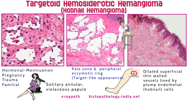
hemangioma is a distinctive cutaneous vascular tumour The histologically descriptive term 'hobnail hemangioma' was proposed to designate these lesions. The commonly proposed etiology is trauma to a preexisting hemangioma. This lesion has also been reported in pregnancy and in a father and son. Hormones may potentiate these tumours. Some authors have suggested a lymphatic origin for hobnail hemangiomas. Age: Usually presents as a single lesion in young and middle aged individuals . Site: The common locations include the lower limb (particularly the thigh) and the trunk, including the shoulder area. Other less common sites are the head, gingiva, and the tongue. Clinical presentation: The lesion may present as angiomatous / pigmented, nontargetoid, flat, or exophytic lesion. The tumour size usually ranges from 0.2 to 3cm in diameter. Some of the tumours have a distinctive targetoid appearance characterized by small violaceous papule at the center surrounded by a pale area and an ecchymotic or brown ring which expands and later disappears. The central papule persists. Microscopic features: This poorly circumscribed vascular lesion consists of a biphasic pattern. Superficial component : Dilated thin walled vessels lined by plump endothelial (hobnail) cells. Fibrin thrombi are seen within some superficial vessels. Some vessels show endoluminal stromal papillae formation. There is no endothelial multilayering or tufting. Deeper component: The vascular spaces are smaller, slit-like and angulated in the deeper dermis. The endothelial cells lining these vessels are less conspicuous. The vessels have a collagen-dissecting, pseudoangiosarcomatous pattern and infiltrated into the dermis and subcutis. There is extravasation of red blood cells and hemosiderin pigments in the surrounding stroma. Cytologic atypia is usually minimal or absent. There is no mitotic activity. Immunohistochemistry: There is variable reactivity of endothelial cells with CD31, CD34, Factor VIII-related antigen and Ulex europaeus agglutinin-1. Smooth muscle actin-positive pericytes are observed focally around some of the abnormal vascular spaces. Differential Diagnosis: Skin changes following radiation therapy ; Dabska's tumour ; Retiform hemangioendothelioma ; patch stage Kaposi's sarcoma ; Well-differentiated Angiosarcoma. Dermatopathology Quiz Case No 61
|
|
|
