Custom Search
|


|
Pathology of Trichilemmoma
|
|
Custom Search
|
|
Pathology of Trichilemmoma
|
| Syn:
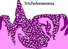
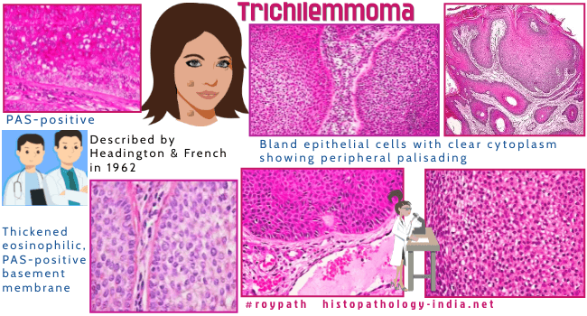
Tricholemmoma. Trichilemmoma arises from the outer root sheath of the hair follicle (mainly of the bulb region). Trichilemmoma was first described by Headington and French in 1962, as a clear cell tumor with differentiation towards the outer hair root sheath. This tumour may be associated with Cowden's syndrome. [ Cowden's syndrome (multiple hamartoma syndrome) is an autosomal dominant disease characterized by multiple cutaneous hamartoma (trichilemmoma, fibroma, verrucous lesions), visceral hamartoma (hyperplastic gastric polyp) or visceral carcinoma (breast carcinoma). ] Clinical presentation : Solitary or multiple papules in adults ; dome shaped, flesh coloured lesions usually less than 5mm in diameter. Sites : Face, nose, eyelids, lips and oral cavity.
Differential diagnosis: Trichilemmal Carcinoma ; Basal cell carcinoma ; squamous cell carcinoma, Hidradenoma; pilar tumour and other adnexal tumours showing focal trichilemmal differentiation. Clear cell poroma (contain duct lumina and there is no basement membrane or peripheral palisading). What is Cowden Syndrome ? [Pathology Infographic]
|
|
Further reading: Headington JT, French AJ. Primary neoplasms of the hair follicle. Histogenesis and classification. Arch Dermatol. 1962;86:430–441. doi: 10.1001/archderm.1962.01590100044012. Trichilemmomas show loss of PTEN in Cowden syndrome but only rarely in sporadic tumors. Trichilemmoma: an immunohistochemical study of cytokeratins. Trichilemmoma of eyelid and eyebrow. A clinicopathologic study of 31 cases. Tricholemmoma. A cutaneous hamartoma. Trichilemmal tumor undergoing specific keratinization: "keratinizing trichilemmoma". |
|
|
Visit:- Infectious Disease Online

Consultant Histopathologist (Kolkata - India)
|
![]()
Copyright © 2022 histopathology-india.net