

Custom Search
|

|
Infectious Disease Online Pathology of Acrodermatitis Chronica Atrophicans Dr Sampurna Roy MD 2023
|

|
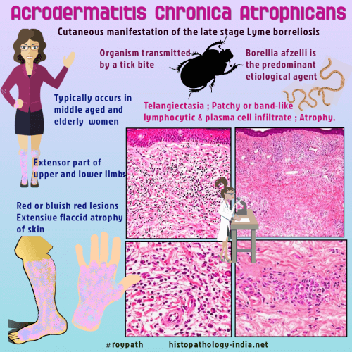
Acrodermatitis chronica atrophicans, the characteristic cutaneous
manifestation of the late stage Lyme borreliosis, typically occurs in elderly
women.
It is a chronic manifestation of infection by Borrelia burgdorferi. Borrelia afzelli is the predominant etiological agent. Acrodermatitis chronica atrophicans was first described by Herxheimer and Hartmann in 1902. Clinical presentation: Early inflammatory stage is characterized by diffuse or localized erythema. It gradually spreads on the extensor surfaces of the extremities and areas around joints. Later there is gradual atrophy of the skin with loss of appendages. Sclerodermatous patches and linear fibrotic bands may be present over ulna and tibia.
Differential diagnosis: Morphea: Acrodermatitis chronica atrophicans shows atrophy of collagen and elastic tissue as well as hypertrophic basophilic elastic tissue; whereas in morphea, sclerosis and polarizing elastic tissue are prominent. Graft-versus-host like reactions may be present in both dermatoses.
|
|
|
Visit:- Dermatopathology Online
 Prof (Dr) Haradhan Roy MD (AIIMS) (1928-2022) (R) Director-Professor and Head of the Dept of Pathology, Calcutta National Medical College, Calcutta University India |

Consultant Histopathologist (Kolkata - India)
|
![]()
Copyright ©
2002-2023 histopathology-india.net