

Custom Search
|

|
Infectious Disease Online Pathology of Adenovirus Infection
|
Adenoviruses cause acute respiratory disease (usually), pneumonia (occasionally), acute follicular conjunctivitis, epidemic keratoconjunctivitis, cystitis, and gastroenteritis (occasionally). In infants, pharyngitis and pharyngeal-conjunctival fever are common. #adenovirus #infection #pathology #histopathology #infectiousdisease #virus |
|
Adenoviruses are nonenveloped, double-stranded DNA viruses associated with
a wide range of clinical syndromes in humans. Adenoviruses typically cause self-limited respiratory, gastrointestinal, or conjunctival disease throughout the year, without significant seasonal variation. Adenovirus infections are most common among children, people living in close quarters (such as college students and military recruits), and immunocompromised patients. Adenoviruses were first isolated in 1953 by Rowe and co-workers from human adenoids removed at surgery. There are 51 immunologically distinct human adenovirus serotypes - (6 species: Human adenovirus A through F). Human adenoviruses have a capsid with icosahedral symmetry. Rodlike structures with knobs at the ends protrude from the capsid. The genome of the virus is a double-stranded DNA linear molecule. When infecting cells in vitro, adenoviruses are capable of lytic infection, latent infection, and transformation. Because of the ability to transform cells and to produce tumours in rodents, they have been considered possible human tumour viruses, but up to now there has been no evidence that links adenoviruses to human tumours.
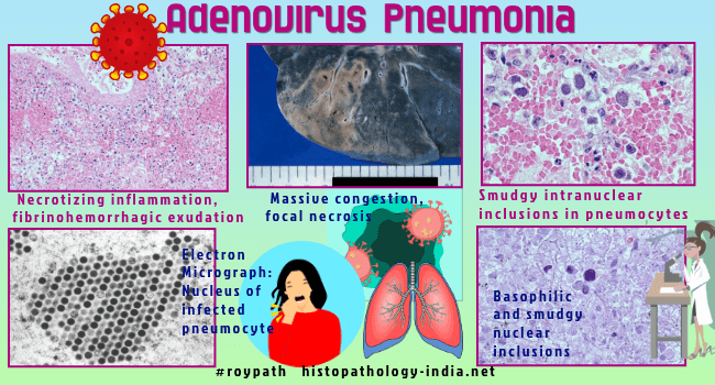
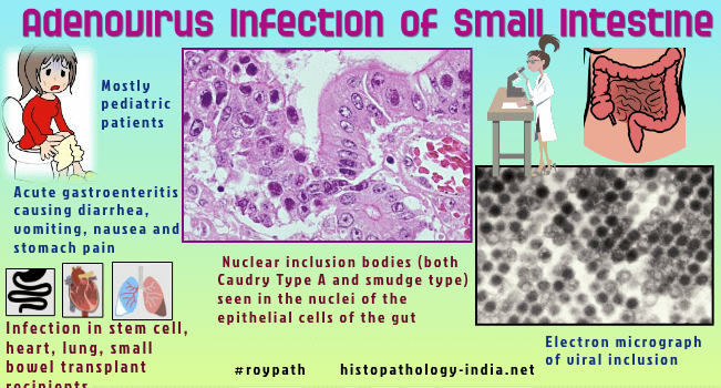
Adenoviruses that are difficult to culture have been associated with 7% to 17% of cases of diarrhea in children. Adenovirus pneumonia is characterized by necrotizing bronchitis and bronchiolitis. There is intense necrosis and desquamation of the respiratory epithelium into the bronchial lumens. These foci of bronchiolitis are surrounded by areas of consolidation, hemorrhage, and atelectasis. At low power the appearance may be confused with the bacterial bronchopneumonia . In the areas of consolidation and among the necrotizing bronchiolar epithelial lesions, cells with intranuclear inclusion bodies may be found. In the early stages cytopathic effects are manifested by granular, slightly enlarged nuclei containing eosinophilic bodies intermixed with clumped basophilic chromatin. The eosinophilic bodies coalesce, forming larges masses to end as a central, granular, ill-defined mass surrounded by a halo. The second type of inclusion, which is more common and probably corresponds to a late-stage infected cell, is designated the "smudge cell". The nucleus is rounded or ovoid, large, and completely occupied by a granular amphophilic to deeply basophilic mass. There is no halo, and the nuclear membrane and nucleus are indistinct. Electron microscopy of the lung demonstrates viral particles in the bronchiolar and alveolar lining cells. Children infected with some strains of adenovirus develop inflammation of the stomach and intestinal tract, which can cause diarrhea and abdominal cramps (gastroenteritis).
|
|
|

![]()
Copyright © 2021 histopathology-india.net