

Custom Search
|

|
Infectious Disease Online
Pathology of Clonorchiasis |
|
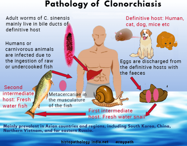
Humans acquire clonorchiasis - infection by Clonorchis Sinensis (Chinese liver fluke), by eating raw or undercooked fish. Adult worms are flat and transparent, live in the bile ducts, and pass eggs to the intestine and the feces. After ingestion by an appropriate snail, the egg hatches in a miracidium. Cercariae escape from the snail and seek out certain fish, which they penetrate and in which they encyst. When human hosts eat the fish the cercariae emerge in the duodenum, enter the common bile duct through the ampulla of Vater, and mature in the distal bile ducts to an adult fluke. The symptoms vary from mild to severe, depending on the number of flukes. An onset with chills and fever indicates bacterial infection from biliary obstruction. Patients with clonorchiasis may die from a variety of complications, including biliary obstruction, bacterial cholangitis, pancreatitis, and cholangiocarcinoma. With massive infection the liver may be up to three times normal size. Dilated bile ducts are seen through the capsule, and the cut surface is punctuated with thick-walled, dilated bile ducts. The flukes, sometimes in the thousands, can be expressed from the bile ducts.
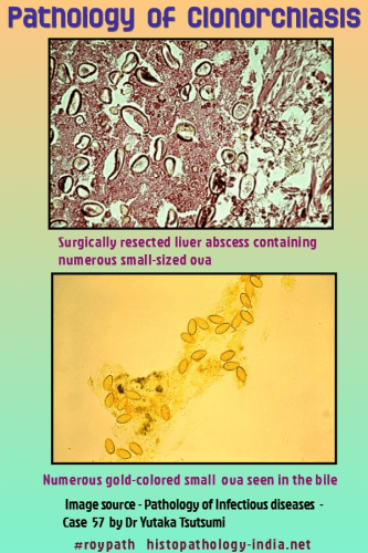
Microscopically, the epithelium lining the ducts is initially hyperplastic and then becomes metaplastic. The surrounding stroma is fibrotic. Secondary bacterial infection is common and associated with pus in the bile ducts. Eggs deposited in the hepatic parenchyma are surrounded by a fibrous and granulomatous reaction. Masses of eggs become lodged in the bile ducts and cause a cholangitis. The pancreatic duct may also be invaded and become dilated, thickened, lined by metaplastic epithelium, and eventually surrounded by scar. The diagnosis is made by identifying the eggs of Clonorchis sinensis in stools or in duodenal aspirates. Treatment is with praziquantel. Cholangiocellular carcinoma originating in either the intra - or extrahepatic bile ducts, is a complication of clonorchiasis. The similar liver fluke endemic in Thailand, Opisthorchis viverrini, also causes cholangiocellular carcinoma.
|
|
|
