|
Histopathology
Images of Colloid Milium
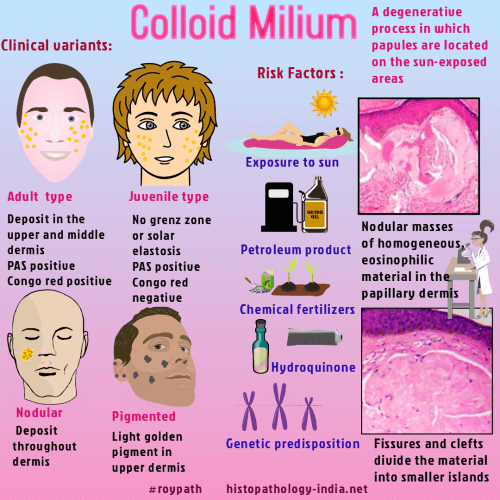
Colloid milium is
an unusual cutaneous disorder characterized by multiple cystic papules.
It is a degenerative process in which the papules are located in the
photoexposed areas.
The material
in the dermis represent a degeneration product of elastic fibers which
is induced by solar radiation.
Colloid milium and
colloid degeneration include at least four distinct clinicopathological
conditions:
- Classic
adult type colloid milium
- Juvenile
colloid milium
- Pigmented
colloid milium (hydroquinone related)
- Colloid
degeneration (paracolloid)
Classic adult type
colloid
milium :
- The adult
form develops in sun-exposed parts of the body in patients who have
actinic-damaged skin.
- Develops in
mid-adult life.
- Plenty of
yellow-brown, semitranslucent papules or plaques ( 1 to 4 mm in
diameter ) are seen in the cheeks, ears, neck, and dorsum of the hands.
- Predisposing
factors are exposure to petroleum products and/or excessive sun
sunlight.
Juvenile type
colloid milium :
-
Exceedingly rare and develop prior to puberty.
- Papules or plaques are seen on the face and neck.
- Some
cases are probably examples of erythropoietic protoporphyria.
Pigmented
type colloid milium :
- Gray to
black papules on the face, following the excessive use of hydroquinone
bleaching cream.
Colloid
degeneration :
- Nodules or
plaque-like areas, on the face and is probably a heterogeneous group.
Histopathology :
Adult form:
- Nodular
masses of homogeneous, eosinophilic material expanding the papillary
dermis and extending into the mid dermis.
- Fissure and
cleft divide this material into smaller islands and fibroblasts are
commonly aligned along the line of fissuring.
- A thin grenz zone of normal collagen, often with elastic fibers (which are also
present between and below the colloid masses), separates the colloid
material from the thinned overlying epidermis.
- Colloid
material may be stained positively with crystal violet and Congo red and
fluorescence with thioflavin T (better result on frozen sections).
- Absence of laminin or type IV collagen differentiates colloid milium from lipoid
proteinosis and primary cutaneous amyloidosis.
Juvenile form:
-
In
most areas grenz zone is absent and basal layer may show hyalinization
with a transition towards the dermal material.
- The colloid
is PAS positive and sometimes, methyl violet positive but it is usually Congo
red negative.
Pigmented form:
- Lightly
pigmented colloid islands are seen in the upper dermis.
Colloid
degeneration (paracolloid):
-
Amorphous, homogenized dermal collagen with less conspicuous clefts,
extends deeply into the dermis.
- The
material is relatively acellular, weakly PAS positive but negative with
Congo red and crystal violet.
Colloid milium: A
histopathologic mimicker of nodular amyloidosis.Arch
Dermatol. 2006
Jun;142(6):784-5.
Ocular Pathology:
Colloid
milium of the upper eyelid margins: a rare presentation.
Ophthalmology. 2002
Oct;109(10):1944-6.
AIM: To
report a rare presentation of colloid milium occurring only on the
upper eyelid margins. DESIGN: Interventional case report. METHODS:
(1) Slit-lamp examination of eyelids and eyes and clinical
examination including the face and a general physical examination;
(2) photography of the lesions on the lids; and (3) excision
biopsy and histopathologic examination. MAIN OUTCOME MEASURES:
Histologic examination confirmed the clinical diagnosis. RESULTS:
Clinical examination and histopathologic findings revealed the
cysts to be colloid milium. CONCLUSIONS: Colloid milium can
involve upper eyelids in isolation, sparing the lower eyelids and
facial skin. Such rare presentations should be kept in mind while
examining similar lesions.
Juvenile
colloid milium associated with ligneous conjunctivitis: report of
a case and review of the literature.
Clin Exp Dermatol. 2000;25(2):138-40.
Juvenile
colloid milium is an extremely rare skin condition with an onset
prior to puberty; it can be distinguished histologically from the
adult form. We report a case of juvenile colloid milium associated
with ligneous conjunctivitis and gingival deposits of an amyloid-like
homogenous eosinophilic material. We hypothesize that all three of
these are the same pathological process occurring at different
sites and review the literature on these associations.
Adult
colloid milium of the eyelid.
Am J Ophthalmol. 1997;123(3):402-3.
PURPOSE:
To report a patient with bilateral upper and lower eyelid margin
nodules that proved to be adult colloid milium. METHODS: After
clinical study, biopsy specimens were obtained and analyzed
histologically and ultrastructurally. RESULTS: Adult colloid
milium can be diagnosed by clinicopathologic correlation.
CONCLUSION: Adult colloid milium should be included in the
differential diagnosis of eyelid margin nodules. |
|
Colloid milium: a review and update.
J Drugs Dermatol. 2007;6(3):293-6.
Colloid
milium (CM) is a rare cutaneous deposition disease with at least 3
distinct subtypes. The exact histogenesis of the condition is
still unresolved and awaits definitive elucidation. Electron
microscopy and immunohistochemical analysis have allowed the
distinction of CM from clinically similar conditions such as
amyloidosis. Successful treatment has been achieved with
dermabrasion and, more recently, with ablative and fractional
laser resurfacing of affected skin.
Pigmented
coalescing papules on the dorsa of the hands: pigmented colloid
milium associated with exogenous ochronosis.J
Dermatol. 2006 Apr;33(4):287-90.
Colloid
milium is a rare cutaneous deposition disorder that frequently
occurs in areas of chronic sun exposure such as the face, neck and
backs of the hands and is characterized by multiple small,
discrete, usually amber-colored, dome-shaped papules that cluster
to form large plaques. A 50-year-old white woman with esophageal
squamous cell carcinoma was referred to us with asymptomatic,
slowly spreading lesions localized to the dorsa of her hands which
had been present for 4 years. The condition was diagnosed
histopathologically as pigmented colloid milium associated with
exogenous ochronosis (EO). Colloid milium associated with EO is
very rarely reported in the published work. We think that a
possible interaction between sunlight and exposure to chemical
fertilizers may have played a role in the pathogenesis of both of
the disorders in our case.
Adult-onset
colloid milium. Presentation of two cases.
Actas Dermosifiliogr.
2005;96(10):674-6.
Colloid
milium is a degenerative process that is characterized clinically
by the development of translucent, yellow, 1-2 mm papules located
in photoexposed areas. Histologically, deposits of a colloid
substance are seen in the papillary dermis. We present two cases
of this infrequent pathology in two male patients, who had been
subjected to intense sun exposure because of their work. We
initiated treatment with photoprotective creams and topical
tretinoin, with little clinical improvement.
Juvenile
colloid milium.
Ann Dermatol Venereol.
2002;129(12):1386-8.
INTRODUCTION:
Colloid Milium is a rare disease. It is characterized by the
development, on sun-exposed areas, of clusters of yellow and
translucid, hard papules, that seeps a gelatinous substance when
opened. This dermatitis predominantly affects elderly patients
and, exceptionally, children. We report a case of juvenile colloid
milium. OBSERVATION: A 14 year-old boy consulted for papular,
translucid or amber-brown lesions, occasionally hemorrhagic on the
cheeks, nose upper lip and the upper edge of the helix of both
ears. He was born to consanguineous parents. Living in a rural
area, he was exposed to sun during agricultural activities. The
lesions had progressed since he was 6 years old and were
exacerbated in the summertime. There was no similar case in the
family. Histological exploration of a papular lesion showed areas
of atrophic epidermis and the presence of a few vacuolized
keratinocytes. The epidermis was lifted by nodular lumps of
amorphous eosinophilic material deposited in the superficial
dermis. Staining with Congo red was negative. DISCUSSION: Juvenile
colloid milium is a benign but unesthetic dermatitis. The inducing
role of sun exposure is obvious, as in colloid milium of adults,
but other pathogenic hypotheses also exist.
Papuloverrucous colloid milium: an occupational variant.
Br J Dermatol.
2000;143(4):884-7.
We report a
case of adult colloid milium in a 47-year-old mechanic with a long
history of professional contact with lubricating oils and of sun
exposure. In addition to the typical translucent papules seen on
the forehead, there were warty papules on the backs of both hands
with unusual histological and ultrastructural features: marked
hyperplasia of the epidermis with orthokeratotic hyperkeratosis
and papillary deposits of colloid material that were contiguous
with the basal layer of the epidermis. The hand lesions caused by
occupational exposure to mineral oils and solar radiation
represent an occupational variant of adult colloid milium.
Adult-type
colloid milium of hands and face successfully treated with
dermabrasion.
South Med J. 1996;89(10):1004-7.
Colloid
milium is an unusual cutaneous disorder characterized by multiple
cystic papules. The adult form develops in sun-exposed parts of
the body in patients who have actinic-damaged skin. We describe
the case of a patient with the classic clinical and histologic
features of this disorder. The patient was successfully treated by
dermabrasion to the hands and affected facial regions and, 10
months later, has not shown any signs of recurrence.
Juvenile
colloid milium: clinical, histological and ultrastructural
features.
J Cutan Pathol. 1992;19(5):434-8.
We report
the clinical, histological and ultrastructural features of
juvenile colloid milium affecting a brother and sister. In this
rare condition, translucent papules develop on sun-exposed areas
of skin, with onset in childhood. Histologically and
ultrastructurally, the papules consist of amyloid-like material
derived from epidermal keratonocytes. A review of the literature
suggests a possible genetic abnormality that leads to sun-induced
degeneration of keratinocytes.
Nodular
colloid degeneration: distinctive histochemical and
ultrastructural features.
Cutis. 1985;36(4):355-8.
We
recently evaluated a patient with a unique form of nodular colloid
degeneration. Lesions consisted of pruritic, purpuric plaques
involving the upper and lower extremities and the neck.
Examination of a biopsy specimen revealed large, fissured masses
of amorphous material in addition to solar elastosis. The
amorphous material showed negative reactions to stains for amyloid,
and other histochemical studies showed results similar to those
for colloid milium. The amorphous masses demonstrated a marked
positive reaction to the Verhoeff-van Gieson stain for elastin.
Electron microscopic examination revealed masses of degenerated
elastic tissue bordered by fibrillar material that resembled
amyloid morphologically. We believe this case represents a nodular
form of solar degeneration with clinical and histologic
resemblances to both colloid milium and cutaneous amyloidosis.
|
|