

Custom Search
|
|
Custom Search
|
|
Dermpath-India Pathology of Elastosis Perforans Serpiginosa Dr Sampurna Roy MD 2022
|
|
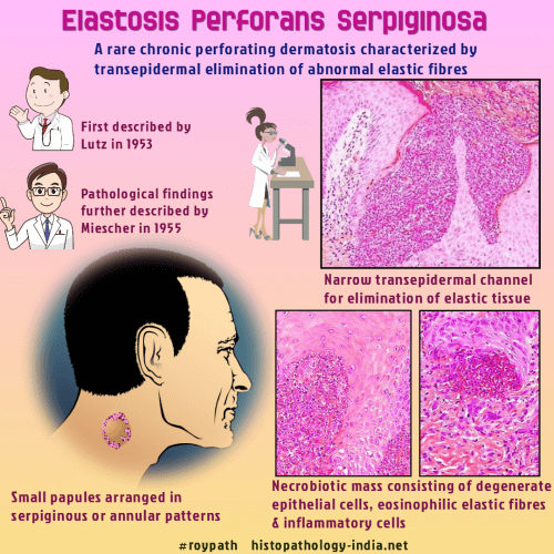
Elastosis perforans serpiginosa
(EPS) is a rare chronic perforating dermatosis which was first
described in 1953 by Lutz. In 1955 Miescher further characterized this
entity on the pathological findings. Elastosis Perforans Serpiginosa is one of four classic primary perforating disorders along with reactive perforating collagenosis, perforating folliculitis, and Kyrle disease. EPS present as asymptomatic to pruritic groups of keratotic papules coalescing in a arcuate serpiginous distribution. It has been reported more commonly in male than female patients and are typically seen in the second decade of life. Face, ear, neck, extremities and trunk are commonly affected. The majority of EPS cases are idiopathic. Some cases are associated with systemic disorder such as Down's Syndrome or disorders of the connective tissue, example, Ehlers-Danlos syndrome, osteogenesis imperfecta, pseudoxanthoma elasticum, or Marfan's syndrome. Rarely it may develop following penicillamine therapy. Important microscopic features are acanthotic, hyperkeratotic epidermis and transepidermal elimination of elastic tissue. Diagnosis of this condition is suspected from hematoxylin-eosin stained sections. It is further confirmed by elastic stain (Verhoeff-Van Gieson stain). Characteristic microscopic picture is the presence of narrow, transepidermal channels which may be straight, wavy or corkscrew shaped. In the lower portion the channels are filled with blue staining necrobiotic masses consisting of mixture of degenerated epithelial cells, inflammatory cells, and brightly eosinophilic degenerated elastic fibers. Elastic stain shows great increase in the amount and size of the elastic fibers in the upper dermis, particularly in the dermal papillae. Differential diagnosis include perforating folliculitis and Kyrle’s disease. These are a group of cutaneous syndromes that are characterized by transepidermal elimination of connective tissue components. However, unlike Elastosis perforans serpiginosa, these lesions do not show increase in elastic tissue in the upper dermis (particularly dermal papillae) on staining with elastic tissue stain.
|
| Reference: Elastosis Perforans Serpiginosa in Association with Scabies Mite |
|
|
![]()
Copyright © 2022 histopathology-india.net


