

Custom Search
|

|
Infectious Disease Online
Pathology of Fascioliasis and Fasciolopsiasis
|
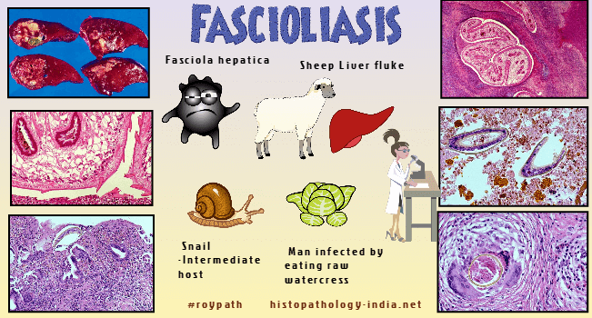
| Fascioliasis is a rare parasitic
infection primarily of the hepatobiliary system caused by one of 2
flatworms, Fasciola hepatica or Fasciola gigantica, which are commonly
referred to as liver flukes. Cattle and sheep are
the most common definitive hosts, but the
flatworms often infect several other grazing animals in the wild. Their
larvae are found on aquatic vegetation. The flatworms most commonly cause
infection in humans when they eat raw aquatic vegetation contaminated with
metacercariae, for example, watercress. Diagnosis is
made by recovering eggs from stools or from the duodenal contents or
bile. Serologic test is an alternative method of confirming early and extrabiliary human fascioliasis. #pathology #histopathology #parasite #infection #Fascioliasis #Fasciolahepatica |
FascioliasisFascioliasis is a rare parasitic infection primarily of the hepatobiliary system caused by one of 2 flatworms, Fasciola hepatica or Fasciola gigantica, which are commonly referred to as liver flukes. F.gigantica lives mainly in tropical climates while F. hepatica is found in temperate climates. They are parasites of many types of herbivorous animals (example - sheep, goats, cattle, buffaloes, horses, rabbits, donkeys and camels). Their larvae are found on aquatic vegetation. Fasciola hepatica is endemic to Europe and Asia, occasionally seen in Northern Africa, Central and South America, and the Middle East, and sporadic cases rarely occur in the United States and the Caribbean. Fasciola gigantica infects domestic livestock across Asia, the Pacific Islands, and some parts of Northern Africa. In parts of Africa and Asia, the 2 species overlap, and their clinical presentations are indistinguishable. Cattle and sheep are the most common definitive hosts, but the flatworms often infect several other grazing animals in the wild. Humans may acquire the infection wherever cattle and sheep industries are prominent. The flatworms most commonly cause infection in humans when they eat raw aquatic vegetation contaminated with metacercariae, for example, watercress. Clinical features and complications - Human fascioliasis can be symptomatic or asymptomatic.More than half of cases are subclinical (asymptomatic), and human infection can be classified as acute or chronic based upon clinical manifestations and laboratory findings.During the acute phase (caused by the migration of the immature fluke through the hepatic parenchyma), manifestations include abdominal pain, hepatomegaly, fever, vomiting, diarrhoea, urticaria and eosinophilia and can last for months. In the chronic phase (caused by the adult fluke within the bile ducts), the symptoms are more discrete and reflect intermittent biliary obstruction and inflammation. Complications include acute hemorrhage from the biliary tree that can present clinically as hematemesis or melena, or aberrant larval migration that can result in ectopic abscesses or nodules in skin, intestine, lung, heart, and brain (cutaneous larva migrans or visceral larva migrans ). Another unusual form of fascioliasis can occur after ingestion of raw Fasciola-infected liver, when flukes surviving mastication attach to the posterior pharynx, thereby causing hemorrhagic nasopharyngitis and dysphagia, known as halzoun in Lebanon and marrara in Sudan. Severe untreated infections may be fatal.
Life cycle - The eggs, passed by the animal in their faeces, require two weeks in fresh water before a miracidium emerges. Miracidia infect a snail (intermediate host) after which infective cercariae emerge from the snail, and encyst on submerged vegetation, such as watercress, that is contaminated by the cysts. Herbivorous mammals (including sheep, goats, cattle, llamas, camels, pigs, deer, and rabbits) become infected by grazing, or indirectly by drinking the surrounding water contaminated with metacercariae. Metacercariae excyst in the duodenum, pass through the wall into the peritoneal cavity, penetrate the liver, and migrate through the hepatic parenchyma into the bile ducts. The larvae mature to adults and live in both the intrahepatic and extrahepatic bile ducts. Later the adult flukes penetrate the wall of the bile ducts and wander back into the liver parenchyma, where they feed on liver cells and deposit their eggs. The eggs lead to abscess formation, followed by a granuloma. The worms induce hyperplasia of the lining epithelium of the bile ducts, portal and periductal fibrosis, proliferation of bile ductules, and varying degrees of biliary obstruction. Diagnosis - Acute hepatitis accompanied by eosinophilia and a history of recent travel or foreign residence suggests the diagnosis of fascioliasis. A history of ingestion of watercress is helpful. Since acute disease occurs before the flukes have begun to lay eggs, stool examinations are negative. Assays for antibody, may be helpful in making the diagnosis of acute fascioliasis, although antibody tests are often negative in early infections. Diagnosis of chronic infections is made through identification in stool of large (130 mm by 60 mm) eggs with poorly defined operculae. Examination of multiple stool specimens using concentration technique should be undertaken since egg output is usually low. Chronic hepatobiliary tract involvement can be assessed with ultrasonography, cholangiography, and computed tomography. Ultrasound may be negative in acute disease, but computed tomography is useful in demonstrating the tracks of migrating larvae in the periphery of the liver. Prevention- Proper cooking or cleaning of aquatic plants before consumption will prevent accidental Fasciola infection.In endemic areas, adequate mass chemotherapy of infected animals and proper sanitary protection of pastures are important control measures. Fasciolopsiasis Fasciolopsiasis, an infection by Fasciolopsis buski, the giant intestinal fluke prevails throughout most of the Orient. Humans, the definitive hosts, acquire fasciolopsiasis by eating uncooked aquatic vegetables contaminated with the encysted cercariae of Fasciolopsis buski . The worm is huge (3 x 7cm) and attaches to the duodenal or jejunal wall. The point of attachment may ulcerate and become infected, causing pain that resembles that of a peptic ulcer. Acute symptoms may be caused by intestinal obstruction or by toxins released by large numbers of worms. The diagnosis is made by identifying the eggs of Fasciolopsis buski, which are similar to those of Fasciola hepatica, in the stool.
|
|
|

![]()
Copyright © 2022 histopathology-india.net