

Custom Search
|


|
Dermpath-India Pathology of Granuloma Faciale Dr Sampurna Roy MD 2022
|
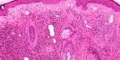
| Granuloma faciale is a rare,
idiopathic dermatosis, of benign course and chronic progression usually
noted in middle aged men. It was described by Wigley in 1945 as eosinophilic granuloma of the skin, and was further defined by Lever and Leeper Pinkus. The lesion presents as erythematous-brownish papules, nodules or plaques, single or multiple, well-delimited and with a smooth surface. There may be accentuation of follicular orifices and superficial telangiectasias. "Peau d'orange" or pitted appearance has been described. The lesions are often asymptomatic and the patient may experience pruritus or a burning sensation. Typical lesion is a solitary plaque on the face. The lesion is usually noted on the sides of the nose, tip of the nose, preauricular area, cheeks, forehead and helix of the ear. There are reports of extra facial and disseminated cases. Extrafacial granuloma faciale has been reported to involve the back, arms, chest, shoulders, and thigh. Possible predisposing factors include actinic exposure, radiation, trauma, allergy, or an Arthus-like reaction. Microscopic features: (i) A grenz zone of uninvolved narrow area of the papillary dermis (not all lesions demonstrate this characteristic finding) (ii) No pathological changes seen in the epidermis. (iii) A dense polymorphous, inflammatory cell infiltrate below the Grenz zone consisting of neutrophils, lymphocytes, histiocytes, eosinophils and histiocytes. (iv) The adnexal structures of the skin are spared. (v) Vascular inflammation, including perivascular inflammation with nuclear dust, damage in the vessel wall and presence of eosinophilic fibrinoid material around the vessels. (vi) Microscopic examination do not show any granulomatous inflammation. Differential diagnosis: (i) Eosinophilic Angiocentric Fibrosis - Although many microscopic features are similar to granuloma faciale, it is located in the sinonasal cavity. (ii) Erythema Elevatum Diutinum - There is no grenz zone. Eosinophilic fibrinoid material is more prominent and related to blood vessels.The number of neutrophils to eosinophils is higher.
|
| Further Reading: Granuloma faciale: a rare disease from a dermoscopy perspective Anais Brasileiros de Dermatologia. 2013;88(6 Suppl 1):97-100. doi:10.1590/abd1806-4841.20132384
|
|
|
