Custom Search
|
|
Custom Search
|
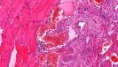
Dermpath-IndiaPathology of Intramuscular HemangiomaDr Sampurna Roy MD 2022
|
 |
| In
1843, Liston first described benign and congenital neoplasms localized
to the muscle of the lower extremity as intramuscular hemangioma. Intramuscular hemangiomas are relatively rare variant of deep-seated hemangiomas that develops within skeletal muscles and accounts for about 1% of all hemangiomas. Age of occurrence of intramuscular hemangioma is usually in the third and fourth decades, but occasionally may present earlier. Intramuscular hemangioma most commonly occurs in the extremities and 14%-21% of intramuscular hemangiomas develop in the head and neck region. The masseter and trapezius muscles are the most common sites in the head and neck, followed by the sternocleidomastoid, mylohyoid, temporalis, and orbital muscles. All patients present with a growing palpable mass which may or may not be accompanied by pain. For the diagnosis of intramuscular hemangioma, plain radiographs, MRI, angiography, and positron emission tomography (PET) may be helpful. The causes of intramuscular hemangioma may be congenital, traumatic, or related to hormone imbalance, but the exact causes are not clear yet. Intramuscular hemangioma can be divided into 3 types, depending on the size of the vessels that constitute the intramuscular hemangioma, and the clinical symptoms and recurrence rates can vary according to the types. (i) Capillary Type Intramuscular hemangioma (ii) Cavernous Type Intramuscular hemangioma (iii) Mixed Type Intramuscular hemangioma The capillary type is the most common among the 3 types, has a relatively short disease duration, is often not accompanied by pain, and has a small size of <10 cm in most cases. The local recurrence rate following surgical treatment of this type is 20%. The best treatment option to date is the complete surgical resection of the tumour. Intramuscular Hemangioma [ Pathology Infographic]
|
|
|
|
|
Visit:- Infectious Disease Online
![]()
Copyright © 2002-2022 histopathology-india.net