

Custom Search
|


|
Dermpath-India Pathology of Lichen Aureus
|
| Lichen
aureus (also called "lichen purpuricus") is a rare and chronic
skin disease.
It is part of the group of pigmented purpuric dermatosis. [Note: Pigmented Purpuric Dermatosis - This group is composed of Schamberg's pigmented purpura, Gourgeot-Blum disease, Kapetanaski disease and Majocchi purpura. These diseases are characterized by a reddish-brown appearance which corresponds histologically to hemosiderin deposition. The lesions present the same histological pattern: a variable degree of lymphocytic infiltrate in the upper dermis associated with hemosiderin deposits. ]
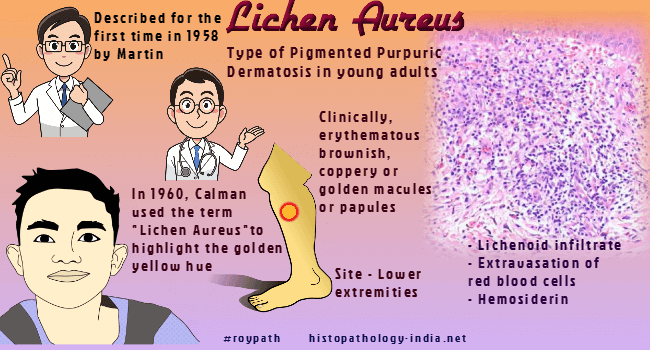
Lichen aureus was described for the first time in 1958 by Martin. In 1960, Calman used the term Lichen aureus to highlight the yellow-golden hue often observed in these lesions. The lesion usually affects young adults and is localized mainly on the lower extremities. It may also involve the forearms and trunk. Most of the lesions are asymptomatic, but there are some reports of itchy or painful lesions. Clinically,. lichen aureus is characterized by an erythematous brownish, coppery or golden macules and/or papules. Linear or segmental presentations have also been described. The onset is sudden, but the course is chronic and can progress slowly or stabilize. Histopathology shows mild or no epidermal alteration. In the dermis there is a typical band-like (lichenoid) inflammatory infiltrate of lymphocytes and histiocytes. There is extravasation of erythrocytes and hemosiderin within histiocytes. Superficial, and deep perivascular lymphocytic infiltrate is present. Perineural and periappendageal infiltrate is exceedingly rare and only case has been reported. The infiltrate, which in half of the cases spares the Grenz zone, tends to be denser than in other pigmented purpuric eruptions, and can be confused with that seen in early stages of mycosis fungoides. There are also fewer extravasated red blood cells than in other pigmented purpuric eruptions.
|
| Further reading:
Lichen aureus: an unusual histopathological presentation: a case report and a review of literature. |
|
|
