

Custom Search
|

|
Infectious Disease Online Pathology of Malakoplakia Dr Sampurna Roy MD 2022
|

|
Custom Search
|
|
Infectious Disease Online Pathology of Malakoplakia Dr Sampurna Roy MD 2022
|
| Malakoplakia
(from the Greek malacos,
soft, and placos, plaques) is a rare granulomatous disease that
occurs commonly in the urinary tract.
It was first described by Michaelis and Gutmann in 1902. Malakoplakia typically involves the genitourinary tract, particularly the bladder, followed by the gastrointestinal tract. Other sites that have rarely been reported include female genital tract, skin, tonsils, middle ears, larynx, lungs, brain, eyes, bone, thyroid, adrenal glands, retroperitoneum, liver, gallbladder, pancreas, and lymph node. Clinically malakoplakia of the bladder shows a female predilection (4:1) with peak incidence in the fifth decade. It tends to present with hematuria, fever, and weight loss and many times is associated with recurrent urinary tract infection. Urine culture usually grows Escherichia coli or other bacteria. Malakoplakia is most commonly seen in immunocompromised patients, such as those on immunosuppressive medications or chemotherapy, and patients with hematopoietic malignancies or diabetes mellitus. Radiation-induced local immunosuppression also poses a risk for malakoplakia, and these cases are easily confused with recurrent malignancies. Although malakoplakia by itself is non-neoplastic,it may be associated with various benign or malignant neoplasms, including lymphoma, adenomatous polyp, and carcinomas of the bladder, prostate, colon and endometrium.
Pathogenesis: 1) Microorganisms might play a role in the pathogenesis. The organisms include Escherichia coli (found in more than two thirds of cases), Mycobacterium tuberculosis, Proteus, and Staphylococcus aureus. Specific bacteria are found in certain categories of patients, for example, coliform bacteria in patients with chemotherapy and Rhodococcus equi in patients with acquired immunodeficiency syndrome. 2) An abnormal or altered immune response has been also implicated in the pathogenesis. 3) The third hypothesis is an abnormal macrophage response because of defective lysosomal function. It is suggested that macrophages in malakoplakia are capable of phagocytosis but unable to digest the bacteria. Partially digested bacteria accumulate in monocytes or macrophages and lead to the deposition of calcium and iron on residual bacterial glycolipid. Gross features: Grossly, malakoplakia can present as soft tan yellow plaques and nodules or even extensive bands. The lesion is usually solitary but can be multiple.
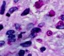
Microscopic features: Histologically, it is defined by sheets of histiocytes called von Hansemann histiocytes or Hansemann cells with accumulation of granular basophilic periodic acid-Schiff - positive, diastase-resistant inclusions and calcified Michaelis-Gutmann bodies, which are pathognomonic but not necessary for diagnosis. The composition of Michaelis-Gutmann bodies has been shown to be 94.6% organic and 5.4% inorganic, with the inorganic components being calcium, phosphorus, and iron. Lymphocytes, plasma cells and neutrophils are interspersed between the histiocytes. Michaelis-Gutmann bodies, which are basophilic targetoid or homogeneous inclusions, 3-10 micrometers in diameter. They are located in the cytoplasm or extracellularly in the stroma. Michaelis-Gutmann bodies can be highlighted by PAS-diastase stain, von Kossa stain for calcium, and Prussian blue stain for iron. Gram stain may show intracellular bacteria. Immunohistochemically, von Hansemann cells are positive for CD68, and negative for cytokeratin, calretinin, and Melan-A.
|
Further reading:Malakoplakia outside the urinary tractGoldblum J, Folpe A, Weiss S. Enzinger & Weiss’ Soft Tissue Tumors, 6th ed: Philadelphia, Elsevier Inc, 2014; 374-6.Rosai J. Rosai and Ackerman’s Surgical Pathology, 10th ed: Philadelphia, Elsevier Inc, 2011; 1251 Malakoplakia of the temporal bone in a nine-month-old infant
|
|
|
Visit:- Dermatopathology Online
![]()
Copyright © 2002-2022 histopathology-india.net
