

Custom Search
|

|
Infectious Disease Online Pathology of Paragonimiasis (Lung Fluke)
|
|
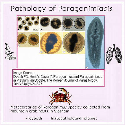
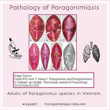
Paragonimiasis is infection by the oriental lung fluke Paragonimus westermani or by one of several other species of Paragonimus. These are the only helminthic parasites of humans that, as adult worms, naturally infect the lungs. Paragonimiasis occurs in Asia, Africa, and South America. Human hosts acquire the infection by eating raw infected crustaceans. Paragonimus eggs are coughed up from the lungs, swallowed and passed in the stool. Miracidia emerge in water and infect a molluscan intermediate host, after which a sporocyst and a generation of redia develop in the mollusk. Infective cercariae emerge and penetrate the gills of a crustacean. The larvae migrate to soft tissue and encyst. After a human host ingests the cyst a metacercaria emerges and penetrates the wall of the stomach, migrates to the diaphragm, bores through the pleura, and settles in the lung, where it matures into an adult worm, which survives for 20 years.
The clinical onset of pulmonary paragonimiasis is insidious, with a diagnostic triad of cough, hemoptysis, and eggs in the sputa or stool. Night sweats, severe chest pain, and pleural effusions are common. Roentgenograms reveal transient diffuse pulmonary infiltrates. Although the adult worms cause pulmonary disease, the larvae occasionally produce lesions at ectopic sites, such as the brain, liver, gut, skeletal muscle, testes, and lymph nodes. The prognosis in pulmonary paragonimiasis is good, but ectopic lesions of brain are often fatal, even with aggressive treatment. The worms provoke leukocytic infiltrates and later, fibrous encapsulation.
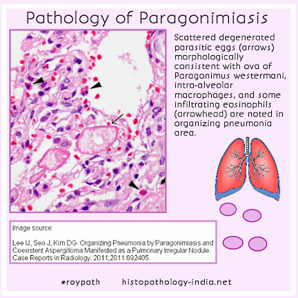
Gravid female worms begin to lay eggs at about 70 days. Cavities that form around the eggs contain worms, eggs and necrotic debris. When these cavities perforate into a bronchiole or bronchus, the eggs are coughed up or become lodged in the lung and provoke fibrosis. Pulmonary paragonimiasis is frequently misdiagnosed as tuberculosis. Eggs in sputa or stools provide the definitive diagnosis. In pleural paragonimiasis, the pleura must be aspirated to obtain eggs. Praziquantel, the drug of choice, is effective against pulmonary paragonimiasis.
|
|
|

![]()
Copyright © 2021 histopathology-india.net