

Custom Search
|


|
Dermpath-India Pathology of Rosacea Dr Sampurna Roy MD 2022
|
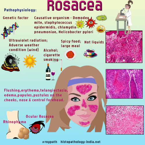
Rosacea (acne rosacea) is an inflammatory disease affecting the central part of face characterized by persistent or recurrent episodes of erythema, papules, pustules and telangiectasias. It is a common disorder of young and middle aged adults. Children are rarely affected. Aetiopathogenesis of rosacea is not clear. Several hypotheses have been documented in the literature and include potential roles for vascular abnormalities, dermal matrix degeneration, environmental factors, and microorganisms such as Demodex folliculorum and Helicobacter pylori. Site: Commonly occurs on the center of the face ( nose, forehead, cheeks, and chin) Gross clinical presentation: The lesion may present red macules, papules, and nodules, as well as pustules and telangiectases. 1. Telangiectatic: Erythematous macules associated with strikingly dilated end blood vessels. 2. Perioral-periocular dermatitis: Papules and pustules around the mouth and eyes 3. Granulomatous: Characterized by tuberculoid granuloma. 4. Rhinophyma: Enlargement of the nose due to proliferation of sebocytes in sebaceous glands. There is formation of infundibular cysts that rupture and elicit granulomatous and fibrosing inflammation 5. Lupus miliaris disseminatus faciei : Papular variant of granulomatous rosacea
Differential diagnosis: Tuberculosis and other mycobacterial infections : Tuberculoid granulomas with caseous necrosis. The lesion is not infundibulocentric. Bacterial infundibulitis and acne vulgaris.
|
|
|
