Custom Search


Dermpath-India

Pathology of Spindle Cell Hemangioma (Haemangioendothelioma)

Dr Sampurna Roy MD                            2022


Dermatopathology Quiz Case: 178

Answer -  Spindle Cell Hemangioma


Spindle cell hemangioma (hemangioendothelioma) is a distinct vascular lesion which was initially considered to be a low grade angiosarcoma when 
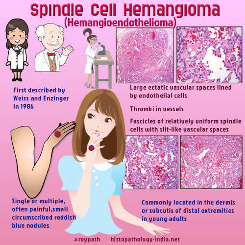
first described in 1986.

Spindle cell hemangioendothelioma. A low-grade angiosarcoma resembling a cavernous hemangioma and Kaposi's sarcoma.

In recent years it has been suggested that spindle cell hemangioma is probably a vascular malformation or the lesion results from  abnormalities of local blood flow.

Spindle cell hemangioma may be associated with anomalies such as Mafucci's syndrome, Klippel-Trenaunay syndrome, lymphedema and early onset varicose vein.

Treatment consists of wide local excision without adjuvant chemotherapy or radiotherapy.
Local recurrence of the lesion is common after incomplete excisions.

Age:  Spindle cell hemangiomas may occur at any age. About 50% patients are 25 years of age or younger.

Clinical presentation: These lesions usually present  as a single or multiple, often painful,  small circumscribed
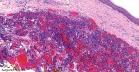
 reddish blue nodules.

Site:  This lesion is usually located in the dermis or subcutis of the distal extremities (particularly the hands ). This lesion can also occur in the proximal extremities, trunk, axilla, ear, vulva, penis, spleen, and pancreas.

Microscopic features:

Microscopically this poorly circumscribed lesion consists of three main components:

- Vascular component displaying gaping thin-walled vessels containing organizing thrombi (features resemble Cavernous hemangioma).

- Solid area of spindle cells together with slit like vascular spaces (features resemble Kaposi's Sarcoma).

- Plump endothelial cells. Some of these cells contain vacuoles or intracytoplasmic lumina.
Bundles of smooth muscle cells are present around the vascular spaces and in the solid areas.

Immunohistochemistry:

Immunohistochemistry reveals that the cells lining the cavernous spaces and the epithelioid cells in the solid area stain positively for endothelial markers CD31 and factor VIII-related antigen.

The spindle cells are immunonegative for endothelial marker CD34 . Most of these spindle cells stain positively for Vimentin and some cells are positive for actin and desmin.

Differential diagnosis:

The main differential diagnosis is with Kaposi's Sarcoma in which cavernous spaces and epithelioid vacuolated cells are usually not present. 

Unlike Spindle cell hemangioendothelioma the spindle cells in Kaposi's  sarcoma stain positively for CD34.


Further reading:


Spindle cell vulvar hemangiomatosis associated with enchondromatosis: a rare variant of Maffucci's syndrome.

Spindle cell hemangioma.

Spindle cell hemangioendothelioma. An analysis of 78 cases with reassessment of its pathogenesis and biologic behavior. 

Multiple enchondromas associated with spindle-cell hemangioendotheliomas. An overlooked variant of Maffucci's syndrome.

Spindle cell hemangioendothelioma: an immunohistochemical and flow cytometric study of six cases.

Spindle cell haemangioendothelioma: probably a benign vascular lesion not a low-grade angiosarcoma. A clinicopathological, ultrastructural and immunohistochemical study.

Spindle cell haemangioendothelioma: a clinicopathological and immuno histochemical study indicative of a non-neoplastic lesion.


Visit:- Infectious Disease Online

Dr Sampurna Roy  MD

Consultant  Histopathologist (Kolkata - India)


Copyright © 2022  histopathology-india.net