Custom Search

Dermpath-India

Pathology of Subcutaneous Panniculitis-Like T-Cell Lymphoma

Dr Sampurna Roy MD                       2022 

Dermatopathology Quiz Case: 174

Answer -  Subcutaneous Panniculitis Like T-Cell Lymphoma


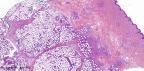
Subcutaneous panniculitis like T-cell lymphoma a rare form of non-Hodgkin lymphoma infiltrating into subcutaneous adipose tissue. It belongs to the group of primary cutaneous T-cell lymphoma.

This is an aggressive tumour and usually affects patients in the 4th decade of life. Some cases have been reported in children.

According to literature review this lesion is more common in female.

Clinically, the lesions present as painful, reddish brown, often ulcerated nodules.

These are usually located on the legs. Cases have been reported in other locations like trunk, arms, axilla and face.

Patient complains of fever, loss of weight and myalgia.

T-cell lymphoma derived from α/β T-cells, is a more aggressive form of lymphoma.


Microscopic Features:

- Histologically, the lesion is characterized by lobular lymphocytic panniculitis.

- The lymphomatous cellular infiltrate is composed of small, medium and large lymphocytes with irregular hyperchromatic nuclei.

- The atypical cells distribute between individual adipose lobules, proliferating and forming a "ring" around adipocytes. In the scanning power the lesion has a lace like-pattern.

- Apoptotic cells and fat necrosis with foamy cells are present in almost all cases.

- "Bean-bag histiocytes" are large phagocytic histiocytes containing cell debris or red blood cells. These are present in many cases.

Note: Cytophagic histiocytic panniculitis is a rare disease. It was first described in 1980. It is characterized by infiltration of subcutaneous adipose tissue by benign-appearing T - lymphocytes and phagocytic histiocytes ("bean bag cells"). Cytophagic histiocytic panniculitis may be an isolated skin disease or associated with non-malignant conditions, such as infections, as well as malignancies, including subcutaneous panniculitis-like T-cell lymphoma.

- Plasma cells are noted in some cases but eosinophils and neutrophils are usually absent.


The cells of Subcutaneous Panniculitis like T-cell lymphoma are T-cells. CD3 is widely used marker since it is never found on B cells.

TCR is also T-cell specific. These are occasionally lost by neoplastic T cells, so that alternative markers such as CD43 or CD45RO, although not specific for T-Cells, may be of supplementary value.

However these antigens are also found in some B cell lymphomas and can be expressed by macrophages and myeloid cells.

CD8 is specific for cytotoxic/suppressor cells. TIA-1, granzyme B, perforin are markers of Cytotoxic T cells.


Further reading:

Subcutaneous Panniculitis-like T-Cell Lymphoma Presenting With Diffuse Cutaneous Edema in a 2-Year-Old Child.

Subcutaneous panniculitis-like T-cell lymphoma in type 1 neurofibromatosis: a case report.

A subcutaneous panniculitis-like T-cell lymphoma mimicking dermatomyositis.

Subcutaneous panniculitis-like T-cell lymphoma: MRI features and literature review.

Subcutaneous panniculitis-like T-cell lymphoma.

Cytophagic histiocytic panniculitis, hemophagocytic lymphohistiocytosis and undetermined autoimmune disorder: reconciling the puzzle.


Visit:- Infectious Disease Online

Dr Sampurna Roy  MD

Consultant  Histopathologist (Kolkata - India)


Copyright © 2022  histopathology-india.net