Custom Search
|


|
Dermpath-India Pathology of Angiolymphoid Hyperplasia with Eosinophilia [Epithelioid Hemangioma] Dr Sampurna Roy MD 2023
|
|
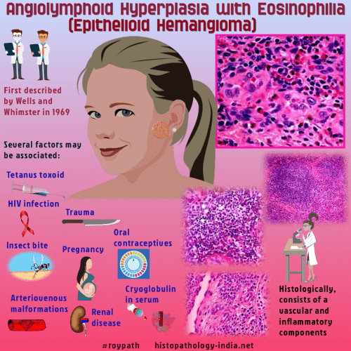
Angiolymphoid
hyperplasia with eosinophilia also known as epithelioid hemangioma is a
distinctive vascular lesion which usually occurs during early and mid
adult life (20 - 40 years). Clinically, this lesion presents as single or multiple pink to red-brown papules and nodules on the face, scalp and ear.
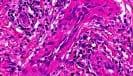
It is still uncertain whether this lesion is a true neoplasm or a
reactive process. The vascular component displays thick and thin walled vessels lined by plump endothelial cells. The epithelioid endothelial cells have rounded nuclei abundant eosinophilic cytoplasm containing occasional vacuoles that represent primitive vascular lumen formation. These endothelial cell protrude into the vessels in a 'tombstone' pattern. Within some thicker vessels there is intravascular proliferations of the endothelial cells.
The inflammatory component
consists of lymphocytes scattered eosinophils and mast cells within the
stroma. Lymphoid follicle may be present.
Differential diagnosis:
Kimura's disease
; epithelioid hemangioendothelioma;
epithelioid angiosarcoma ;
injection site granuloma (epithelioid
cells are not present). Kimura's disease
is a deeper lesion and reactive lymphoid follicles and are present
together with a dense infiltration of eosinophils sometimes forming
eosinophilic abscesses.
|
|
|