Gastrointestinal Stromal Tumour


http://www.histopathology-india.net/VascularTumours.htm

               HISTOPATHOLOGY INDIA.COM

                               Path Quiz Case-7    

       Diagnosis:   Fibrous Hamartoma of Infancy

        Dr Sampurna Roy MD

 
June 2008
Surgical-Pathology.com

Histopathology-India.net

Pathology-India.com

Pancreatic Pathology Online

Gall Bladder Pathology Online

Paediatric Pathology Online

Paraganglioma-Online

Endocrine Pathology Online

Eye Pathology Online

Ear Pathology Online

Cardiac Path Online

Lung Tumour-Online

Mesothelioma-Online

Pulmonary Pathology Online

Nutritional Pathology Online

Environmental Pathology Online

Pathology Quiz Online

Dermpath-India

GI Path Online

Soft Tissue Pathology

Case Index

Infectious Disease Online; INDEX: A-D ; INDEX: E-L ; INDEX: M-P INDEX: Q-Z ; FUNGAL DISEASE ; VIRAL DISEASE.

E-book - History of  Medicine with special reference to India

Vascular tumours

Angiokeratoma

Epithelioid hemangioma (angiolymphoid hyperplasia with eosinophilia)

Lobular capillary hemangioma (pyogenic granuloma

  - Bacillary angiomatosis

  - Verruga Peruana

Masson's Tumour (Intravascular papillary endothelial hyperplasia)

Acro-angiodermatitis / pseudo-Kaposi's sarcoma

Reactive angioendotheliomatosis

Infantile Hemangioma

Glomeruloid hemangioma 

Acquired tufted angioma

Verrucous hemangioma

Cherry angioma/senile angioma

Arteriovenous hemangioma 

Targetoid hemosiderotic hemangioma(Hobnail hemangioma)

Spindle cell  hemangioma / hemangioendothelioma

Kaposiform hemangioendothelioma

Retiform hemangioendothelioma

Papillary intralymphatic angioendothelioma (Dabska's tumour)

Composite hemangioendothelioma

Kaposi's sarcoma

Epithelioid hemangioendothelioma

Angiosarcoma

Glomus tumour

Hemangiopericytoma

Angiolipoma

Aggressive angiomyxoma

Angiomyofibroblastoma

Angioleiomyoma

Angiomyolipoma

Dermatofibroma (aneurysmal variant)

Spindle cell lipoma (Angiomatoid variant)

Kimura's disease

       DermPath-India

       Site created by

   Dr Sampurna Roy MD


http://www.histopathology-india.net/dermpath.htm


Acrodermatitis chronica atrophicans

Actinomycosis

Adenovirus

African Histoplasmosis (Histoplasma Duboisii)

AIDS: Cutaneous lesion associated with AIDS

AIDS related malignant tumour

African Trypanosomiasis

Alphaviruses causing Encephalitis

Amebic Meningoencephalitis

American Trypanosomiasis

Amoebiasis

Ancylostomiasis

Angiostrongyliasis

Anisakiasis

Anthrax Infection

Arenavirus

   Path Case 7 :Case history and images:


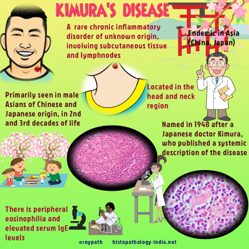
Kimura's disease is a rare, benign chronic inflammatory condition which commonly occurs among the Oriental population.

The exact cause of this disease is unknown but it is believed to result from an immunological reaction that is allergic or autoimmune in nature.

The lesion shows no evidence of malignant transformation.  Recurrence may occur following surgical excision.

Although the Asians are commonly affected some cases have been reported in the Caucasians and in the African population.

Age:  Usually occurs in the second and third decade of life.

Site:  Commonly located in the head and neck region particularly in the pre or post auricular region and rarely in the oral mucosa, orbit, and the scalp.

Rarely, Kimura's disease may occur in the parotid or other salivary glands.

Other rare sites include limbs and trunk, vulva, spermatid cord, inguinal and axillary lymph nodes and peripheral nerves.

Clinical presentation:  The patient  presents with painless subcutaneous nodules or plaques.

Lymphadenopathy is present in more than 50% cases.

Kimura's disease is often associated with peripheral blood eosinophilia, raised ESR and serum IgE.

Accurate diagnosis of Kimura's disease is based on clinical and histological findings.


Microscopic features: Image Link1 ; Image Link2 ; Image Link3 ; Image Link4 ; Image Link5.

Features of the skin lesion :

1. Lymphoid follicles with prominent germinal centre.

2. Fibrous tissue surrounding the lymphoid follicles.

3. Inflammatory cellular infiltrate in the interfollicular area consisting of small lymphocytes, plasma cells and eosinophils. In areas, scattered eosinophilic microabscesses are identified.

4. Thin-walled blood vessels are increased in number lined by flat or cuboidal endothelial cells.

Features of the involved lymph node:

1. Marked hyperplasia of the germinal centre. Germinal centres are often  well vascularized and contain eosinophilic deposits and polykaryocytes (Warthin-Finkeldey type).

2. Extensive eosinophilic infiltration consisting of mature eosinophils in follicular and interfollicular areas and occasional eosinophilic microabscess formation.

3. Proliferation of thin-walled blood vessels in the paracortex.

4. Increase in the number of plasma cells and mast cells in the paracortex.

Differential diagnosis:

Angiolymphoid hyperplasia with eosinophilia :

Previously, angiolymphoid hyperplasia with eosinophilia and Kimura's disease were regarded as the same lesion.  But these conditions are now regarded as two separate entities.

1. Multiple, small  well circumscribed dermal or subcutaneous papules or nodules with well-defined borders. (In Kimura's the lesion is located in the deep soft tissue or the subcutis without significant change of the overlying skin initially ).

2. Less frequently associated with lymphadenopathy and peripheral blood eosinophilia.

3. Histologically, unlike Kimura's disease the blood vessels are lined by plump epithelioid like endothelial cells. These cells demonstrate large nucleus and abundant eosinophilic cytoplasm. Prominent cytoplasmic vacuoles are present.

4. Dense lymphocyic infiltration is present in only some cases of ALHE.

 
ABSTRACT

Kimura T et al. Unusual granulation combined with hyperplastic changes of lymphatic  tissue. Trans Soc Pathol Jpn. 1948;37: 179-180

Kimura disease: a clinicopathologic study of 21 cases.
Am J Surg Pathol. 2004;28(4):505-13.

Refractory hypertension and anemia in end-stage renal disease: an unusual manifestation of Kimura's disease.
Ren Fail. 2003 May;25(3):499-507.

Kimura's disease with unusual eosinophilic epithelioid granulomatous reaction: a finding possibly related to eosinophil apoptosis. Hum Pathol 2002;33(5):561-4

Kimura’s Disease: A Diagnostic Challenge .PEDIATRICS Vol. 110 No. 3 September 2002, pp. e39

Angiolymphoid hyperplasia with eosinophilia should not be confused with Kimura's disease. Acta Ophthalmol. Scand 2001;79: 91-93

Polymerase chain reaction (PCR) for human herpesvirus 8 and heteroduplex PCR for clonality assessment in angiolymphoid hyperplasia with eosinophilia and Kimura's disease. J Cutan Pathol 2001;28(7):363-7

Kimura's disease in a chronic hemodialysis patient. Am J Nephrol 2001;21(1):47-50

Detection of Epstein-Barr virus DNA in a patient with Kimura's disease. Int J Dermatol 2000;39(8):618-2.

Steroid-sensitive anemia in a boy on dialysis--an association with Kimura disease.Pediatr Nephrol 2000;15(3-4):183-5

Kimura's disease and angiolymphoid lymphoid hyperplasia with eosinophilia: clinical and histopathological differences. J Am Acad Dermatol 1992; 27: 954-958 

Kimura's disease; clinical, histological and immuno- histochemical studies.  Acta Med Okayama. 1992; 46: 449-455

Kimura's disease. Involvement of regional lymph nodes and distinction from angiolymphoid hyperplasia with eosinophilia. Am J Surg Pathol. 1988;12: 843-854

Lymphadenopathy of Kimura's disease. Am J Surg Pathol. 1988 ;12: 843-854.

Your Banner
PULMONARY PATHOLOGY:

Congenital Cystic Adenomatoid  Malformation ; Acute Respiratory Distress Syndrome  ;Sarcoidosis ;Bronchiolitis ; Emphysema ; Bronchial Asthma ;Chronic Bronchitis Pulmonary Alveolar Proteinosis ; Lipid Pneumonia ; Pulmonary Hypertension ;Pulmonary edema ;Pulmonary Infection ; Pneumococcal Pneumonia ; Haemophilus influenza Infection;Klebsiella Pneumoniae ; Mycoplasma Pneumonia ; Pneumocystis Pneumonia ; Legionellosis ; Localized Fibrous Tumour of the Pleura ; Biphasic Epithelial/Mesenchymal Lung Tumours ; Pulmonary Carcinosarcoma ;Pulmonary Blastoma ; Large Cell Neuroendocrine tumour;

Myxoid Tumours of Soft Tissue

Classification of Soft Tissue Tumour

Gross examination of soft tissue specimen          

A practical approach to histopathological reporting of soft tissue tumours

Grading of soft tissue tumours

Lipomatous tumours

Neural tumours

Myogenic tumours

Fibroblastic/Myofibroblastic tumours

Myofibroblastic tumours

Fibrohistiocytic tumours

ChondroOsseous tumours

Soft TissueTumours of Uncertain Differentiation               

Notochordal Tumour - Chordoma

Extra-adrenal Paraganglioma

Gastrointestinal Stromal Tumour

               
FUNCTIONAL ANATOMY OF THE HEART

ANATOMY OF THE ATRIUM

ANATOMY OF THE VENTRICLE

ANATOMY OF THE CORONARY ARTERIES

AUTOPSY EXAM. OF CORONARY ARTERIES

EXAMINATION  OF CARDIAC  VALVES

CARDIAC  VALVE  DISEASE

MITRAL  VALVE LESIONS

PULMONARY VALVE DISEASE

TRICUSPID VALVE DISEASE

CARDIOMYOPATHY

CONGESTIVE HEART FAILURE

congenital heart disease

Ischemic heart disease    

Angina pectoris

Myocardial infarction                
 
hypertensive heart disease  
 
RHEUMATIC FEVER AND RHEUMATIC HEART DISEASE
 
PATHOLOGY OF ASCHOFF BODIES OR NODULES
 
myocardiTIS
 
GIANT CELL MYOCARDITIS    

pericardial disease  

INFECTIVE ENDOCARDITIS

CARDIAC HEMOCHROMATOSIS

CARDIAC AMYLOIDOSIS

HISTOPATHOLOGY REPORTING OF PERICARDIAL SPECIMEN

HEART TRANSPLANTS - PATHOLOGICAL EXAMINATION

ENDOMYOCARDIAL BIOPSY-(ALLOGRAFT REJECTION):

ISHLT SYSTEM FOR GRADING REJECTION

POST-OPERATIVE CARDIAC PATHOLOGY

PERIOPERATIVE CARDIAC PATHOLOGY

PRIMARY TUMOURS OF THE HEART

REPORTING OF CARDIAC TUMOURS

CARDIAC MYXOMA

CARDIAC RHABDOMYOMA

PAPILLARY FIBROELASTOMA

CARDIAC FIBROMA

CARDIAC LIPOMA

CARDIAC HEMANGIOMA

CARDIAC TERATOMA

MESOTHELIOMA OF ATRIOVENTRICULAR NODE

PURKINJE CELL TUMOUR

CARDIAC PARAGANGLIOMA

MALIGNANT TUMOURS OF THE HEART

CARDIAC LYMPHOMA